


Radiography and ultrasound are at the core of any equine veterinary practice with a sports horse caseload. As both technology and, subsequently, our clinical understanding of the intricacies of the pathophysiology of equine lameness has developed, there is an increasing requirement and even expectation to perform advanced imaging in order to obtain a definitive diagnosis.
Assessing the Risk
Choosing to perform advanced imaging as part of a lameness diagnostic workup depends on the modality and equipment available. Part of the decision-making process, therefore, requires a balanced risk assessment of whether or not to perform general anaesthesia to obtain images.
The inherent risks of subjecting even a healthy equine patient to general anaesthesia are well-documented. However, factors such as owner compliance, time proximity to competition, increased clinic costs, and technician resources might mean imaging under general anaesthesia is not practical, regardless of the health risks.
So, what if there is an alternative? Recently, we have seen the emergence of several standing limb CT systems on the market. Considering they all aim to achieve the same goal, the difference in approach to solving the problem of imaging the standing horse varies quite dramatically.
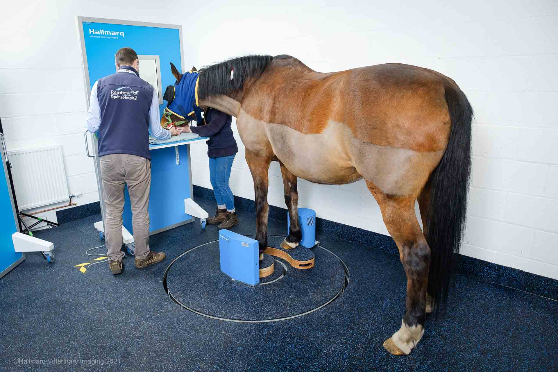
Safety First
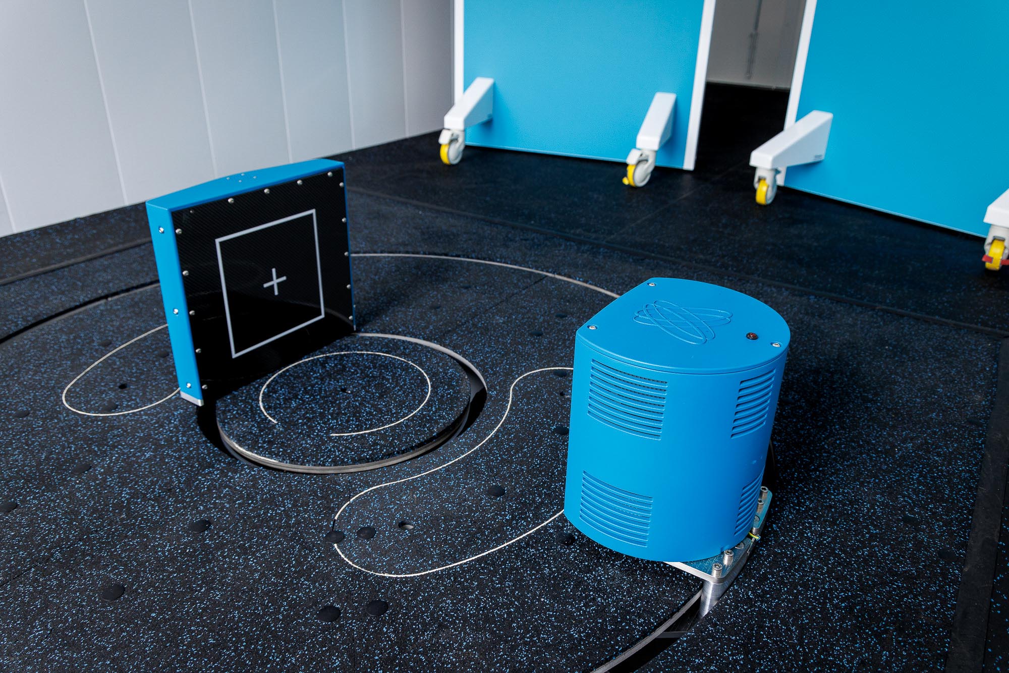
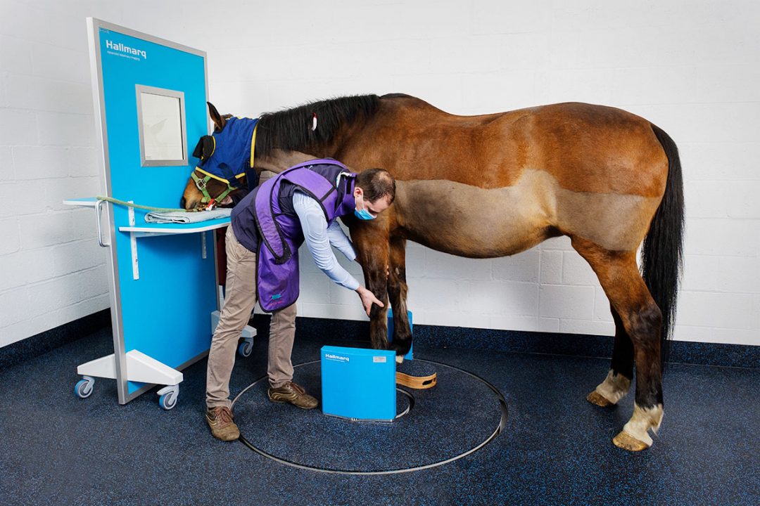
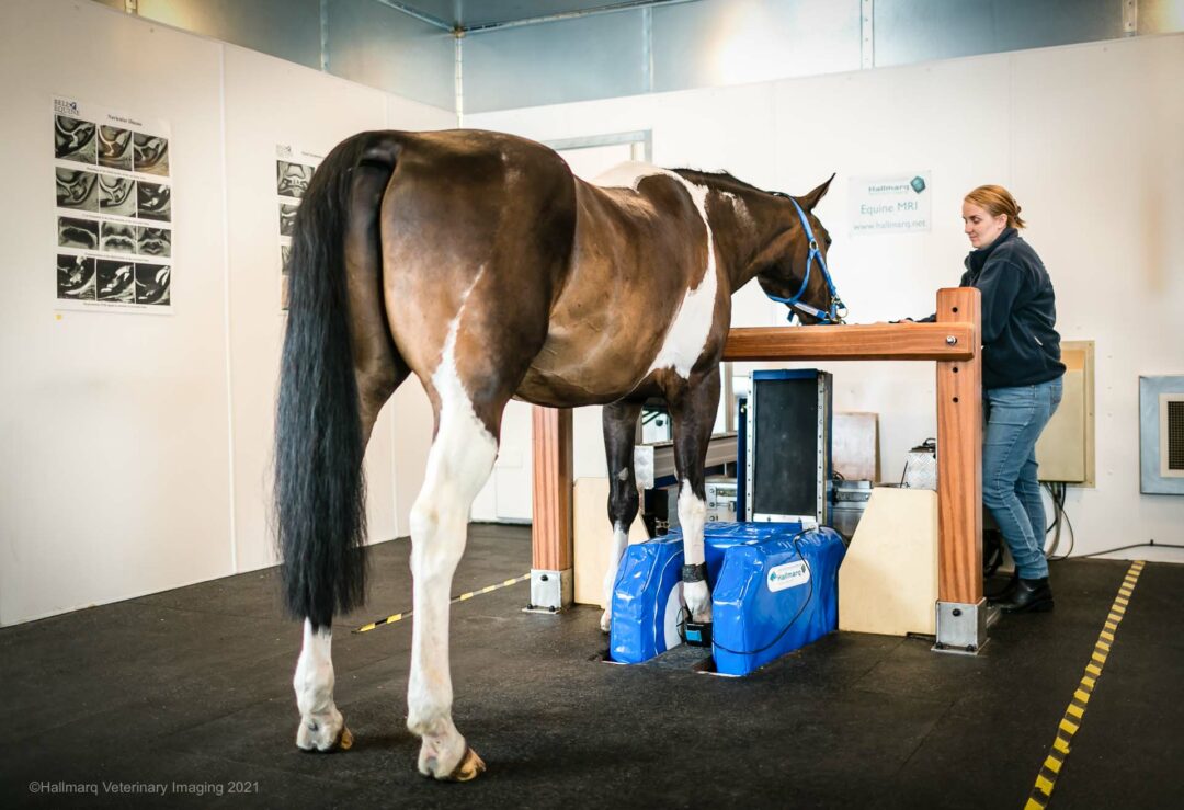
World-renowned for the development of the Standing Equine MRI system, Hallmarq Veterinary Imaging has now launched its Standing Equine leg CT machine (slCT). With the detector and generator at floor level in this walk-in, walk-out system, cone beam technology revolves around the limb to capture 3D image sets in 1 minute. This unique machine is supported by 20 years of experience in motion correction reconstruction software, to deliver superior 3D images. With patient safety paramount, the Hallmarq slCT does not restrict the limb of the horse in any way. It is specifically designed for easy and immediate emergency exit by the patient and handler should the need arise.
The Benefits of Cone Beam Technology
Although cone-beam technology is not widely used in veterinary medicine currently, it offers many additional benefits when imaging the distal limb of the horse. In addition to returning high-quality diagnostic images, this technology is relatively low-cost when compared to fan-beam CT. Furthermore, the operator radiation risk is comparable to widely used digital radiography. The Hallmarq system allows the horse handler and technician to remain in the room with the patient at all times, shielded by a lead screen and ensuring safety for both.
Safe, Effective, Affordable 3D Imaging
The Hallmarq slCT system is designed for ease of use. It enables fast imaging of the patient, typically taking around 15 minutes to complete an examination. Images are sent directly to your image-reading and storage device. This safe, effective, affordable product could help you and your clinic take lameness diagnosis to another level, offering an accessible solution for almost every equine practice.
Article previously published in The Horse