

When it comes to equine lameness diagnosis, acquiring the best possible MRI images starts way before the patient enters the MRI room. In this article, we revisit the basics to look at what good MRI technique means for the horse, owner, and veterinarian alike.
Preparation, Preparation, Preparation
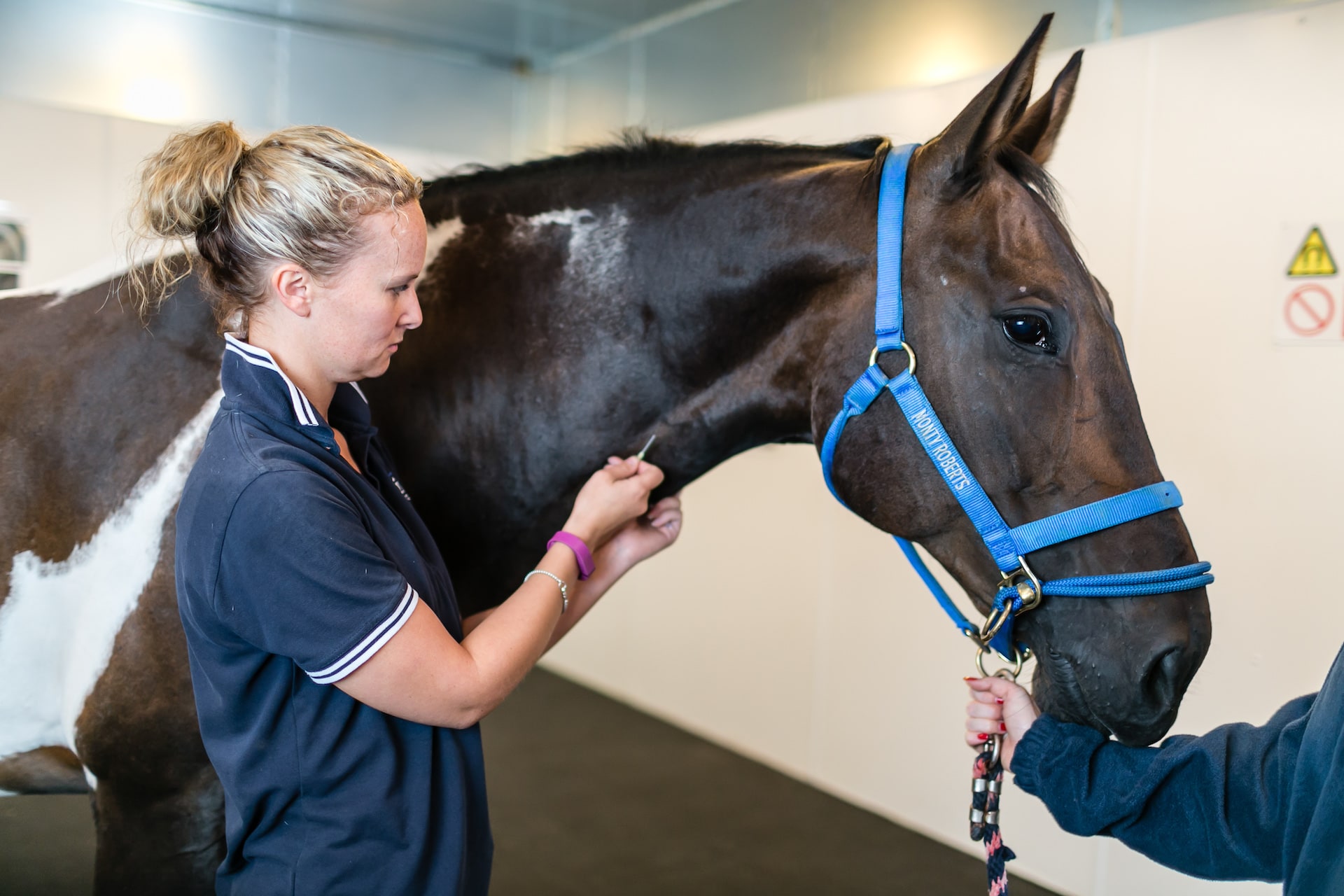
Ensuring that your patient is as relaxed as possible before the procedure will pay dividends. The visiting horse should be given time to stand in the box or stall, urinate and settle down prior to going into the scan room.
The connection between horse and image is invaluable and looking at the lameness pattern on the day of the scan is beneficial to the final outcome. It is also important to see and evaluate the feet. Information on the shoe, trimming, balance, and any distortion to the coronary band will help you interpret everything you see on the MRI.
Most practices take one or two x-rays prior to performing the MRI to help check for clench fragments which can cause artefacts on the images. Time spent on preparation of the feet before the scan is performed will reap rewards.
Sedation techniques vary from practice to practice, but their use and administration is vital. A gently sedated horse will stand better throughout the scan process. Once the horse is in the scan room, the end goal is a comprehensive set of high-quality sequences taken from the region of interest and in a reasonable time.
There are many factors that affect what is achievable and all of which are dependent on the individual horse. It’s important therefore to tailor the scan for each patient.
The Right Sequence
Sequence selection is case-specific and it’s important to know why, when, and how to use them. Firstly, a basic set of pulse sequences needs to be acquired by the MRI Operator:
- T1 for example is designed for anatomical structures
- T2* and STIR look at fluids and where they should not be.
- T2 weighted and fast-spin echo provide excellent contrast for looking at the ligamentous tissues within the foot.
A combination of all these sequences, in multiple planes, allows plenty of room to interpret the images alongside the standard set required. Much is dependent on available time and the skill of the operator in their acquisition.
The equine foot is very complicated and it’s important to evaluate as many structures as possible. The dynamic process that MRI enables is key to diagnosis. There are specific areas where anatomic detail is required:
- Dorsal margin lesion on the DDFT
- Distal border fragments on the navicular bone
- The flexor cortex to see fine areas of fluid accumulation signalling early pathology
- Coffin joint cartilage and fibre cartilage on the back of the navicular bone
There is a difference between a radiology report and logical interpretation. Information about the foot, nerve blocks used, lameness history, and the MRI images all need to be considered before prognosis and diagnosis. For example, reporting on Condylar fractures is far more helpful when the broader context is taken into account.
The more information and the better the communication with the referring vet then the more useful the comprehensive report.
Communication, Communication, Communication
The procedure either succeeds or fails with how we talk to the client. Communication, as always, is key to their understanding and expectations. Advance information on what to expect before they bring the patient to the clinic benefits both parties. Knowing that their horse may need to stay overnight, for example, helps with their pre-planning.
Despite MRI being the gold standard in lameness diagnosis, it is not infallible, and 100% sensitivity and specificity is not possible. But, with MRI proving diagnostic in over 90% of lameness cases, its efficacy is not to be ignored.
MRI provides the best technique available to the equine vet under “reasonable” conditions Additionally, without the need for general anaesthetic the procedure is risk-free to the horse and therefore less expensive for owner and vet.
Your client will undoubtedly ask lots of questions such as is it navicular syndrome, osteoarthritis, a ligament injury, or tendon injury? However, be careful not to over-complicate the advice given; it’s a fine balance between too much and too little information.
Clinically Correct
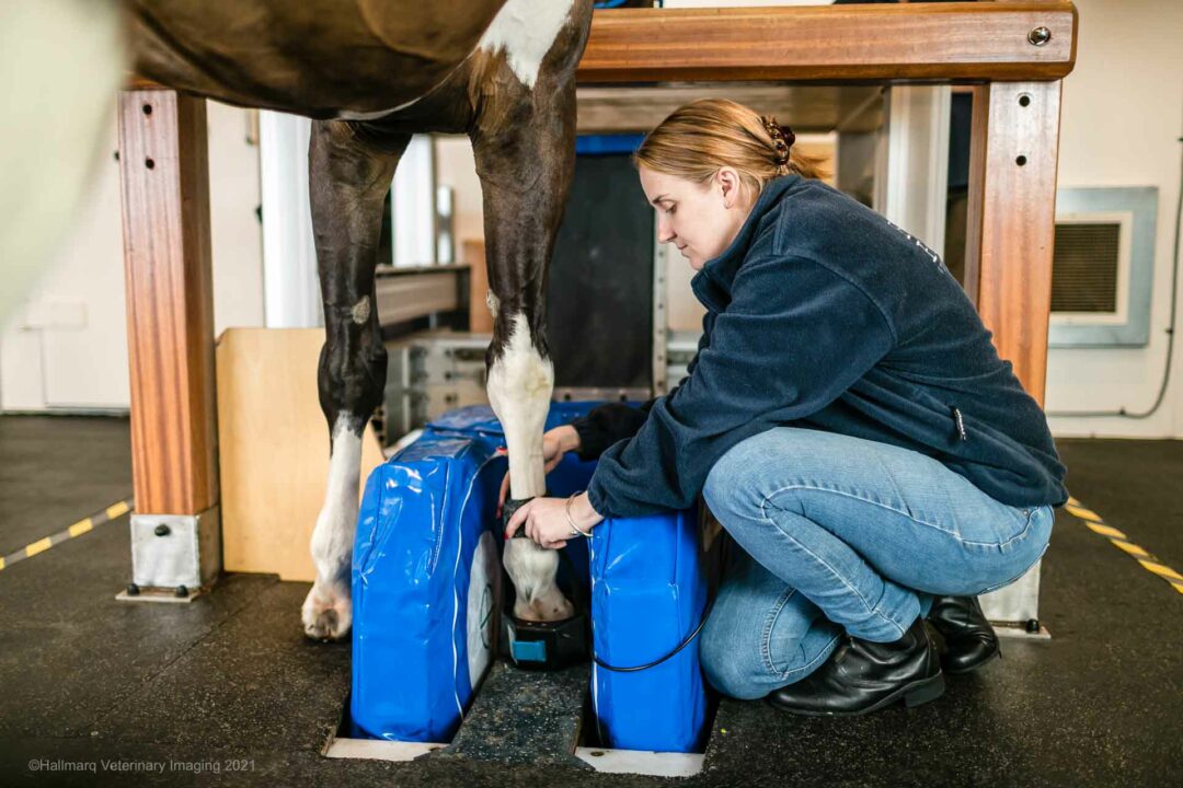
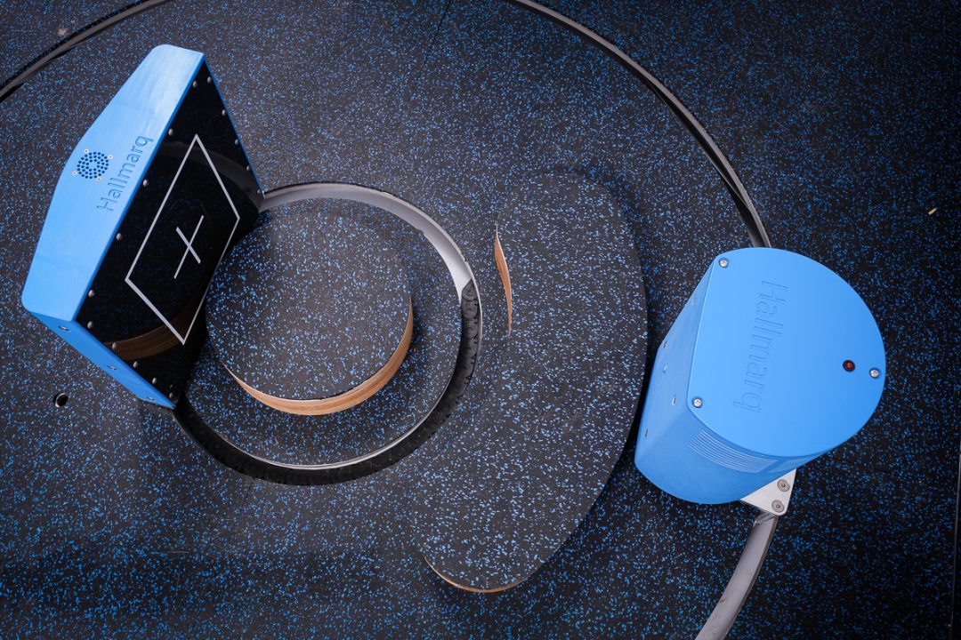
Whilst Hallmarq’s unique Standing Equine MRI machine and award-winning motion correction software helps reduce artefact created by the horse’s natural sway, good horse handling makes all the difference.
Work towards slice thickness being as thin as is achievable to get as much resolution as possible. Remember, the field of view is smaller than a high-field system or CT scan, so be realistic about where you’re looking and what it is you’re looking for.
Hallmarq has worked hard to optimize images of the distal limb, cartilage imaging, and the potential for follow-up without exorbitant costs. This means that preventative scanning close to competition for horses in training is not only feasible but highly beneficial.
What’s the Take Home?
When it comes to good MRI technique in the equine patient, it’s clear that time spent with both horse and owner before the scan is time well spent. Good protocol, an awareness of the broader context and open and honest communication with the referring vet and horse owner will reap rewards for everyone involved in MRI image acquisition.
Taken from a series of talks given by Sarah Powell MRCVS, at a meeting held by Rainbow Equine Hospital
Click here to access the full series on Hallmarq’s YouTube Channel.
For more on the Standing Equine MRI process click here