


The onset of a head tilt is a seemingly innocuous yet potentially indicative sign of underlying neurological issues. Fortunately, magnetic resonance imaging (MRI) plays a pivotal role in understanding the causes of head tilt and guiding accurate treatment for our pets. Hallmarq’s Small Animal Medical Director, Dr. Simon Platt, helps shed some light on this condition and how MRI can help aid diagnosis.
Head tilt – cause for concern?
While a persistent tilt of the head to one side may initially appear benign, it can signify a range of conditions affecting the vestibular system – a complex network responsible for balance and spatial orientation. From benign peripheral vestibular disorders to more sinister central nervous system lesions, the underlying cause of the head tilt demands careful investigation to ensure timely intervention and optimal outcomes for our patients.
Unlike traditional radiography or computed tomography (CT), MRI offers unparalleled detail and contrast resolution, allowing for exquisite visualisation of soft tissues within the brain and spinal cord. This non-invasive technique utilises powerful magnetic fields to generate detailed cross-sectional images, enabling veterinarians to evaluate the vestibular system with unprecedented clarity.
MRI – the diagnostic cornerstone
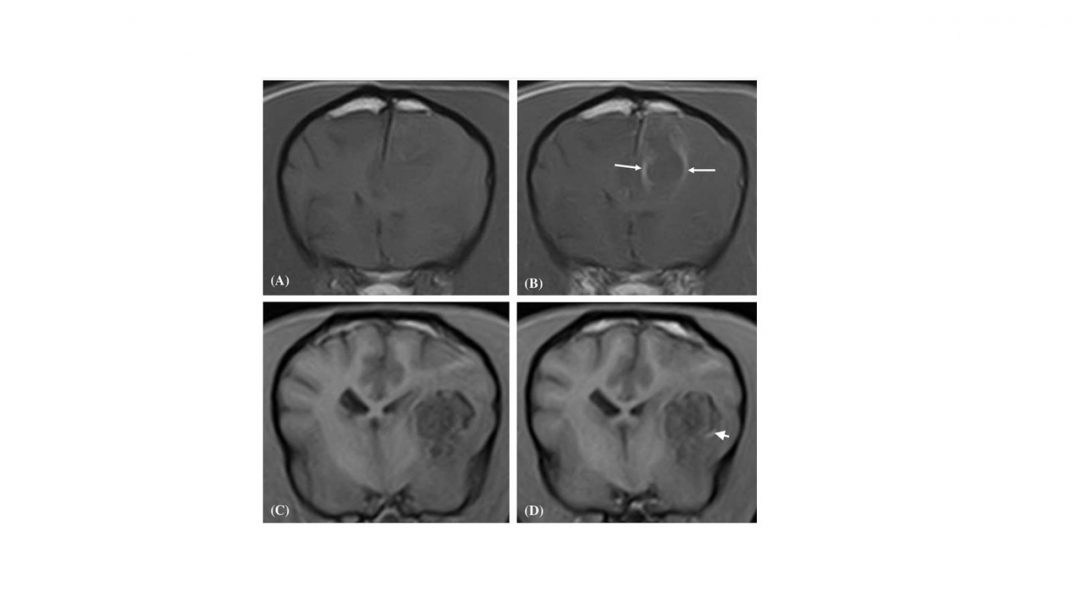
When confronted with a pet presenting with a head tilt, MRI provides a cornerstone in our diagnostic arsenal. By obtaining high-resolution images of the brain and surrounding structures, we can pinpoint the exact location and nature of any abnormalities contributing to the clinical signs. Whether it be a tumour compressing the cranial nerves, an inflammatory lesion affecting the inner ear or the brainstem, or even a stroke affecting the cerebellum, MRI provides invaluable insights that help shape our treatment approach and prognosis.
Moreover, MRI plays a crucial role in ruling out differential diagnoses and guiding therapeutic decision-making. By differentiating between peripheral and central vestibular pathology, we can tailor our treatment strategies accordingly, whether it involves medical management for inflammatory conditions, surgical excision of tumours, or supportive care for idiopathic disorders.
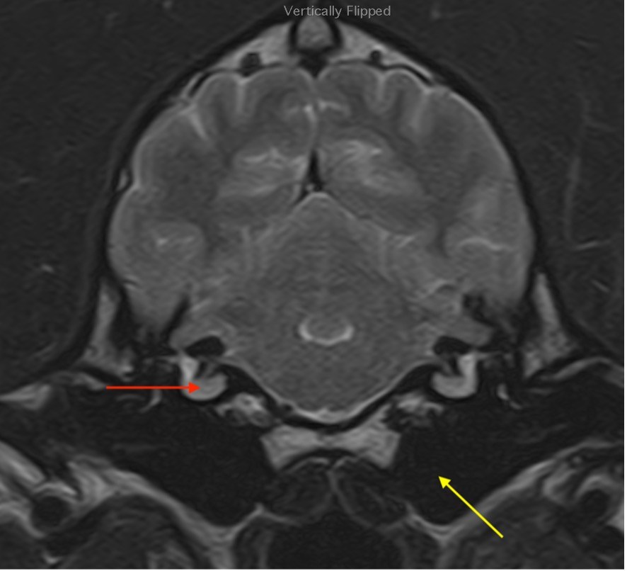
Peripheral diseases include
- idiopathic dysfunction, which is essentially diagnosed by ruling other diseases out and MRI is integral for that purpose. It is a self-limiting disease and rarely recurs
- inner ear infections (otitis interna)
- polyps
- external and middle ear neoplasias
All of those listed here can be identified with reasonable specificity on MRI.
Central diseases include:
- stroke which is again specifically identified with this form of advanced imaging
- inflammatory disease (meningitis/encephalitis)
- neoplasia
The presence of these latter two conditions may be difficult to distinguish between. However, by default, their location rules out a peripheral condition. Additionally, the safety of other tests that may be helpful – such as CSF – can also be determined using MRI which can assess whether there is a potential for elevated intracranial pressure and as such would be a contra-indication for such diagnostics.
In conclusion:
The head tilt may be a perplexing puzzle but, with MRI, we can understand the specific location of the disease and gain a more educated insight into its cause allowing more accurate therapeutic advice as well as prognostication.
INTERESTED IN VISIONARY VETERINARY IMAGING?