

Haemorrhage in the brain of dogs can be due to several causes which include coagulation problems of a primary or secondary nature, neoplasia and inflammation. The confirmation of an intracranial haemorrhage requires cross-sectional imaging such as MRI. However, sometimes haemorrhage can be difficult to confirm using imaging and can be confused with other diseases such as brain tumours.
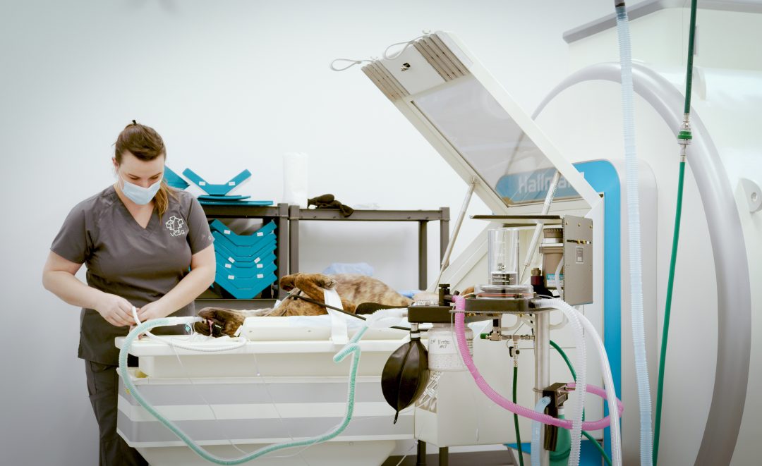
This study describes the characteristics of intracranial haemorrhage when identified using a 1.5T MRI which helps to improve diagnostic confidence. Some of the images for this study were acquired using a Hallmarq magnet, serving to support the role of Hallmarq’s award-winning technology in clinical research.
MRI Findings Aid Treatment and Prognosis
The differentiation of solitary intra-axial hematomas from hemorrhagic neoplasms based on their magnetic resonance imaging (MRI) features is challenging. The treatment and prognosis for these two disease entities are vastly different and the distinction between them is often based on MRI findings alone. The aim of this study was to describe the 1.5 tesla MRI features of canine intra-axial hematomas and correlate these findings with the evolution of haemorrhages described in human brains. Retrospective evaluation of patient details, clinical signs, and MRI findings of dogs with intra-axial hematomas that were histopathologically confirmed or determined via repeat MRI study and/or the resolution of neurological signs.
Inclusion Criteria is Paramount
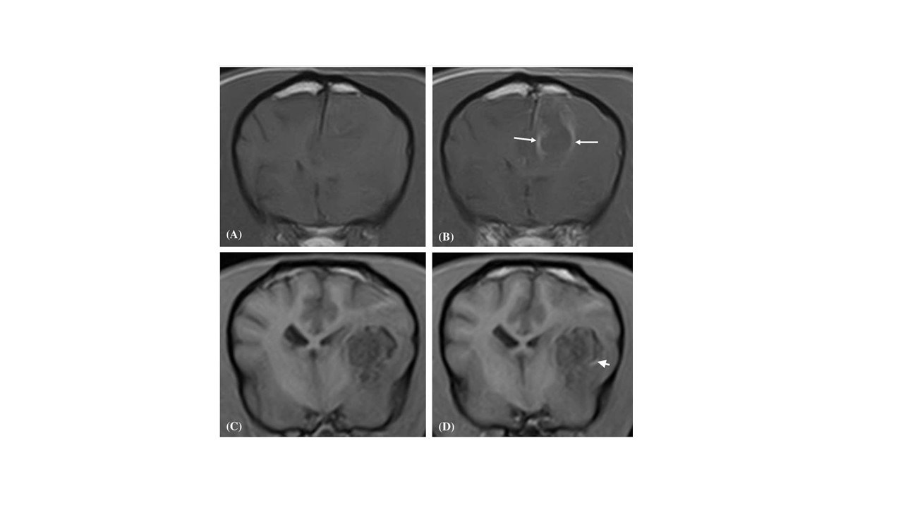
Ten dogs met the inclusion criteria. All 10 hematoma lesions were determined to be 2–7 days in age. On MRI, all 10 hemorrhagic lesions were comprised of two distinct regions; a relatively thin T1-weighted (T1W), T2-weighted (T2W) and gradient echo (GRE) hypointense (9/10) peripheral border region and a large central region that was heterogenous but predominantly T1W, T2W and GRE hyperintense (8/10). The peripheral border region was complete in its integrity in all 10 cases on T2W and GRE sequences. Contrast enhancement was present in (6/10) hematoma lesions and was always peripheral in nature with no evidence of central enhancement associated with any of the lesions. An intra-axial hematoma should be suspected in solitary hemorrhagic space-occupying lesions that have a complete hypointense peripheral rim, elicit a peripheral contrast enhancement pattern, and display the expected temporal pattern of hematoma evolution.
References:
Whitlock J, Holdsworth A, Morales C, Garosi L, Carrera I. 1.5 Tesla Magnetic Resonance Imaging Features of Canine Intracranial Intra-axial Hematomas. Front Vet Sci. 2021 Dec 24;8:778320. doi: 10.3389/fvets.2021.778320. PMID: 35004926; PMCID: PMC8739912.