Magnetic Resonance Imaging (MRI) is a powerful diagnostic tool that can provide detailed images of a dog’s internal structures, making it invaluable in investigating cases of neck pain. When a dog presents with neck pain, it is crucial to accurately diagnose the underlying cause to ensure effective treatment. Hallmarq’s Small Animal Medical Director, Dr. Simon Platt, helps shed some light on this condition and how MRI can help aid diagnosis.
Is MRI the only option?
MRI can offer detailed insights that other imaging modalities, such as radiographs or CT scans, might miss. However, it is important to remember that the less expensive and more available imaging option of neck radiographs (X-rays) can help to identify several common abnormalities. These include bone infections (osteomyelitis / discospondylitis), trauma (fracture / luxations), congenital vertebral malformations, and neoplasia of the vertebral bone. With limited soft tissue detail, however, the extent and effect of these diseases on the spinal cord cannot realistically be confirmed.
Common causes of neck pain in dogs
Neck pain in dogs can stem from various causes, including:
- Intervertebral Disc Disease (IVDD): A common condition where the discs between the vertebrae of the spine degenerate and can rupture or extrude, causing tremendous damage to the spinal cord and significant ongoing cord compression.
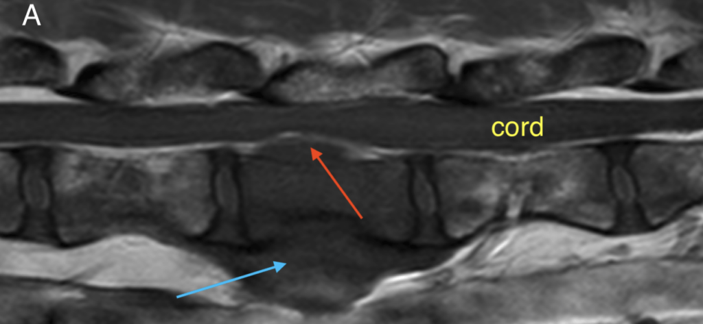
- Spinal Cord Tumors: Tumors within or around the spinal cord can cause pain and neurological deficits (Fig 1A and Fig 1B below). The most common tumors affect the vertebral column surrounding the cord, with less common tumors affecting the nerves leaving the cord and the cord itself.
- Infections and Inflammation: Conditions like meningitis, be it infectious or presumed immune-mediated in origin, can cause severe neck pain, as can infections affecting the vertebrae.
- Congenital Abnormalities: Some dogs are born with abnormalities of the vertebral column. Sadly, these can lead to pain and neurological dysfunction as they mature. Malformations of the cord itself (syringomyelia) are also common in certain breeds and can be responsible for intermittent discomfort.
- Trauma: Injuries from accidents can cause fractures, luxations, and spinal cord hemorrhage, all of which can require immediate attention to improve the chances of patient recovery.

The role of MRI in diagnosis
- Detailed Imaging of Soft Tissues: MRI excels at imaging soft tissues, including the spinal cord, meninges, nerve roots, intervertebral discs, and surrounding muscles. This level of detail can help identify issues like disc herniation, inflammation, or tumors that might not be visible on X-rays or CT scans.
- Early Detection of Diseases: MRI can detect diseases of the bone such as tumors and infection, before they become apparent on other imaging modalities. This allows for earlier intervention and treatment, which can improve the prognosis.
- Differentiating Between Conditions: MRI can help differentiate between various causes of neck pain and assess their impact on the spinal cord. This is crucial for determining the appropriate treatment plan particularly when a surgical decision needs to be considered.
- Guiding Treatment Plans: The detailed images provided by MRI can help veterinarians plan surgeries or other treatments with greater precision. For instance, in cases of IVDD, knowing the exact location and extent of disc herniation can help in planning a surgical approach and ensuring that the true extent of the disease is known, prior to operating.
Conclusion
MRI can provide detailed images of the soft tissues of the neck, the vertebral column, and the spinal cord enabling accurate diagnosis and effective treatment planning. For dogs with neck pain, an MRI scan can be the key to identifying the specific cause enabling a more precise and effective treatment.
INTERESTED IN VISIONARY VETERINARY IMAGING?