


Cerebral Microbleeds
A 14-year-old, female spayed Pug dog presented with seizures. Otherwise considered healthy, the dog underwent MRI using the Hallmarq 2nd generation 1.5T unit.
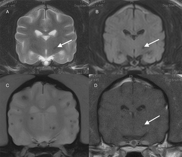
(A) T2W, (B) FLAIR, (C) T2* GRE and (D) T1W post-contrast images identify multiple, well defined punctate intra-parenchymal lesions, the majority of which are at the grey-white matter junctions. They are of homogenously T2W and T1W hypointense without notable enhancement and are seen as signal voids on T2* images. The lesions, which are all less than 5mm in diameter, are barely seen on most sequences with the most significant one being identified within the left thalamus (A, B and D arrow), but they are easily visible on T2*, emphasizing the essential need for this sequence on all brain imaging studies.
In this dog, the imaging appearance of the multiple signal voids is compatible with intraparenchymal haemorrhages. Hemorrhagic lesions less than 5mm in size are called cerebral microbleeds (CMBs) and are often incidental.
With thanks to Dr Christianne Massicotte DVM, DACVIM (Neurology) and the team at Veterinary Care & Speciality Group, Chattanooga, TN, for providing this case study.


