History
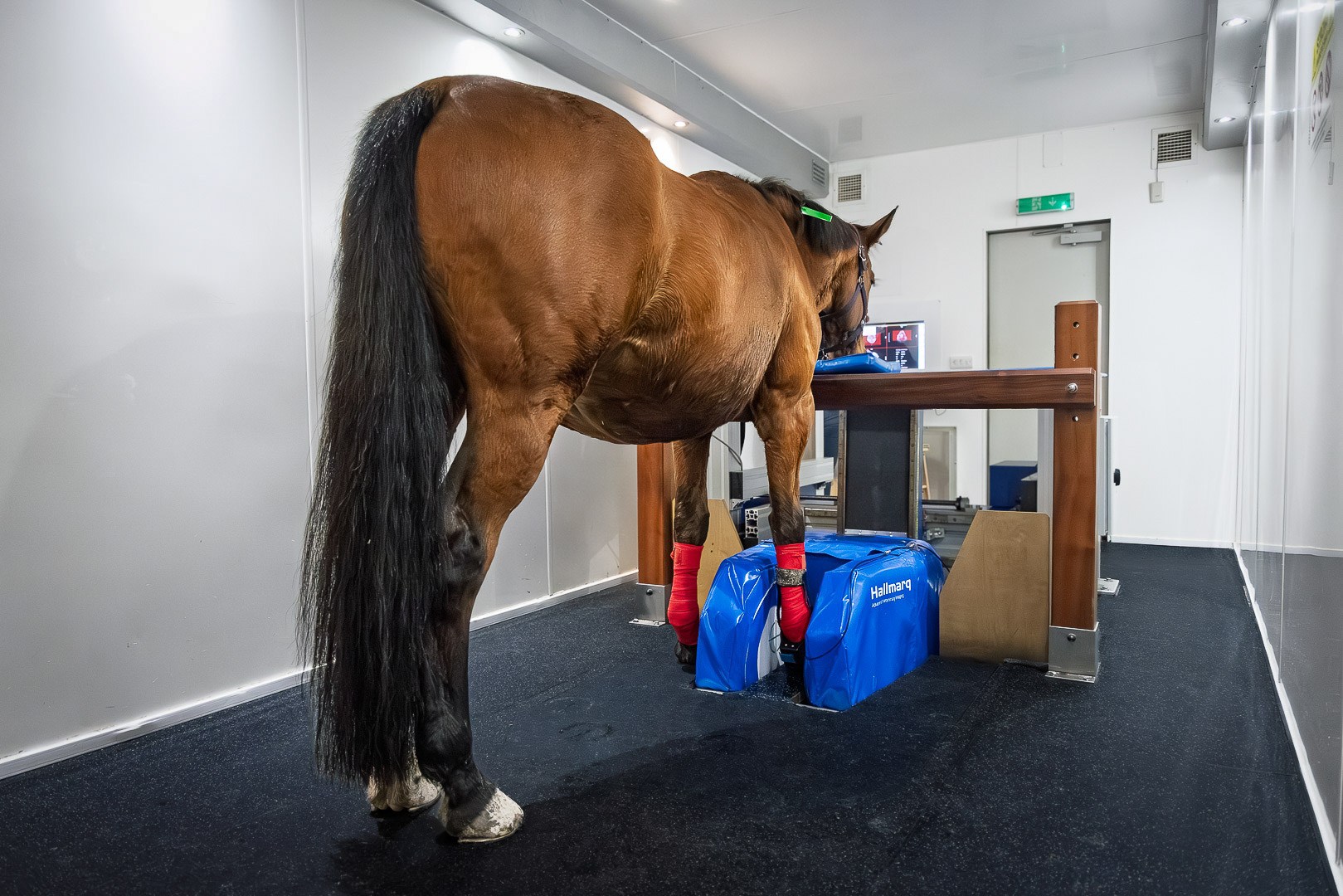
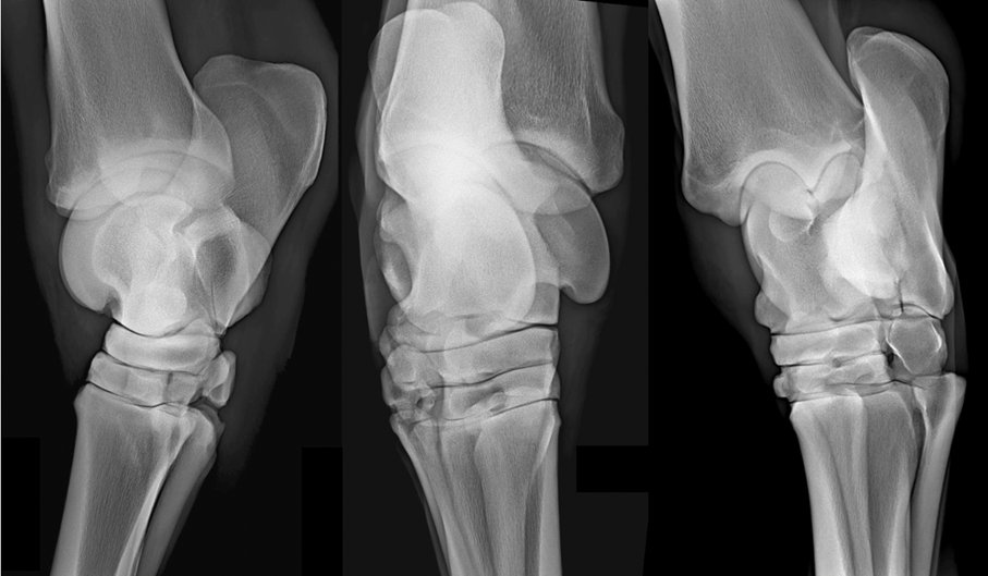
A 10 year-old showjumper was examined for acute left hindlimb lameness with effusion of the tarsocrural joint. Radiographs of the left tarsus showed abnormal sclerosis of the central tarsal bone. Based on the history and clinical signs, fracture of this bone was highly suspected, but not clearly visible on radiographs.
MRI findings
Examination of the left tarsus, with Standing Equine MRI, confirmed an incomplete, non-displaced, bi-articular fracture of the central tarsal bone, dorsomedial-plantarolaterally oriented.
The plantar half of the central tarsal bone was surrounded by severe oedema-like signal, centered on the fracture plane, indicative of marked inflammation. Sclerosis was present on the dorsal aspect of the central tarsal bone, more pronounced dorso-medially.
Transverse images of the left tarsus were obtained with iNAV enhanced motion correction software applied (Figs 2A and 2B). iNAV proves particularly beneficial when imaging the fetlock, tarsus, carpus, or high suspensory delivering improved detail and the clarity required for accurate assessments.
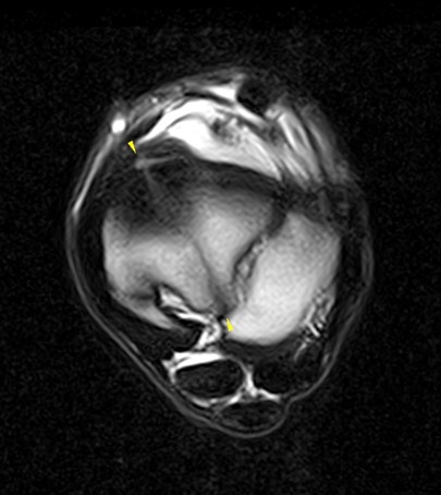
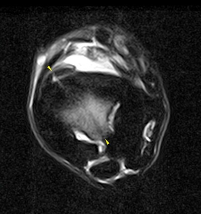
Fig. 2A and 2B: (above) T2 FSE iNAV and STIR FSE iNAV transverse images of the left tarsus showing the fracture plane extending dorsomedially-plantarolaterally within the central tarsal bone. There is a severe increased STIR FSE (bone oedema-like signal) in the plantar half of the central tarsal bone, centered on the fracture plane.
Standing MRI has considerably improved our assessment of lameness cases. In addition, the introduction of iNAV allows us to obtain images of exceptional diagnostic quality even for difficult areas, such as the tarsal region. In the present case, the fracture line and the area of bone oedema-like signal are clearly defined with minimal motion artifacts, which would not have been possible otherwise.”
Dr Claudia Fraschetto DMV, DECVSMR, ISELP Cert, Clinique de Grosbois
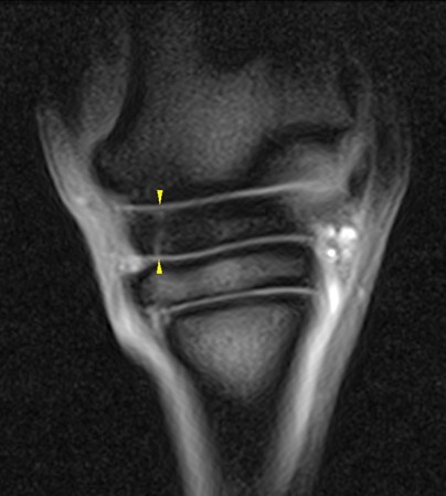
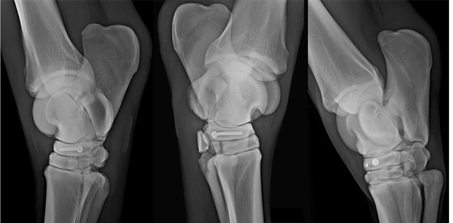
Thanks to the comprehensive MRI study, surgical fixation of the fracture was then successfully performed.
Conclusion
The horse was discharged from the hospital a few days later with an appropriate rehabilitation programme.
With thanks to Dr Claudia Fraschetto DMV, DECVSMR, ISELP Cert, and the team at Clinique de Grosbois, France, for providing us with this case study.

