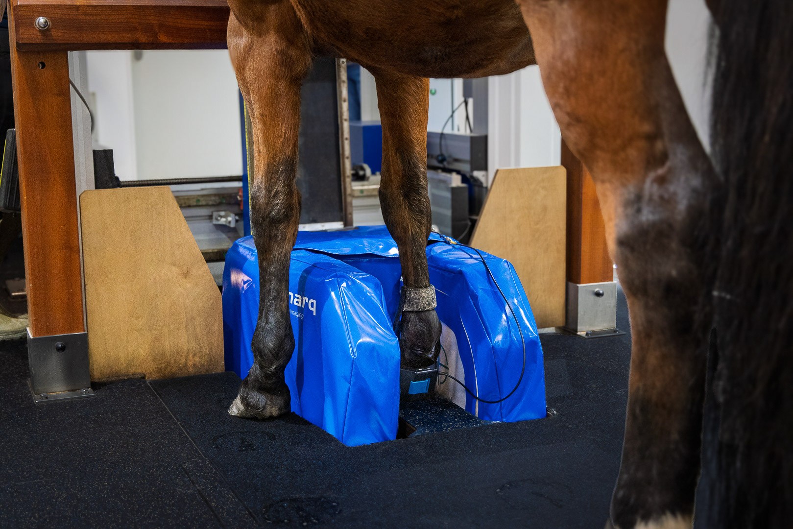
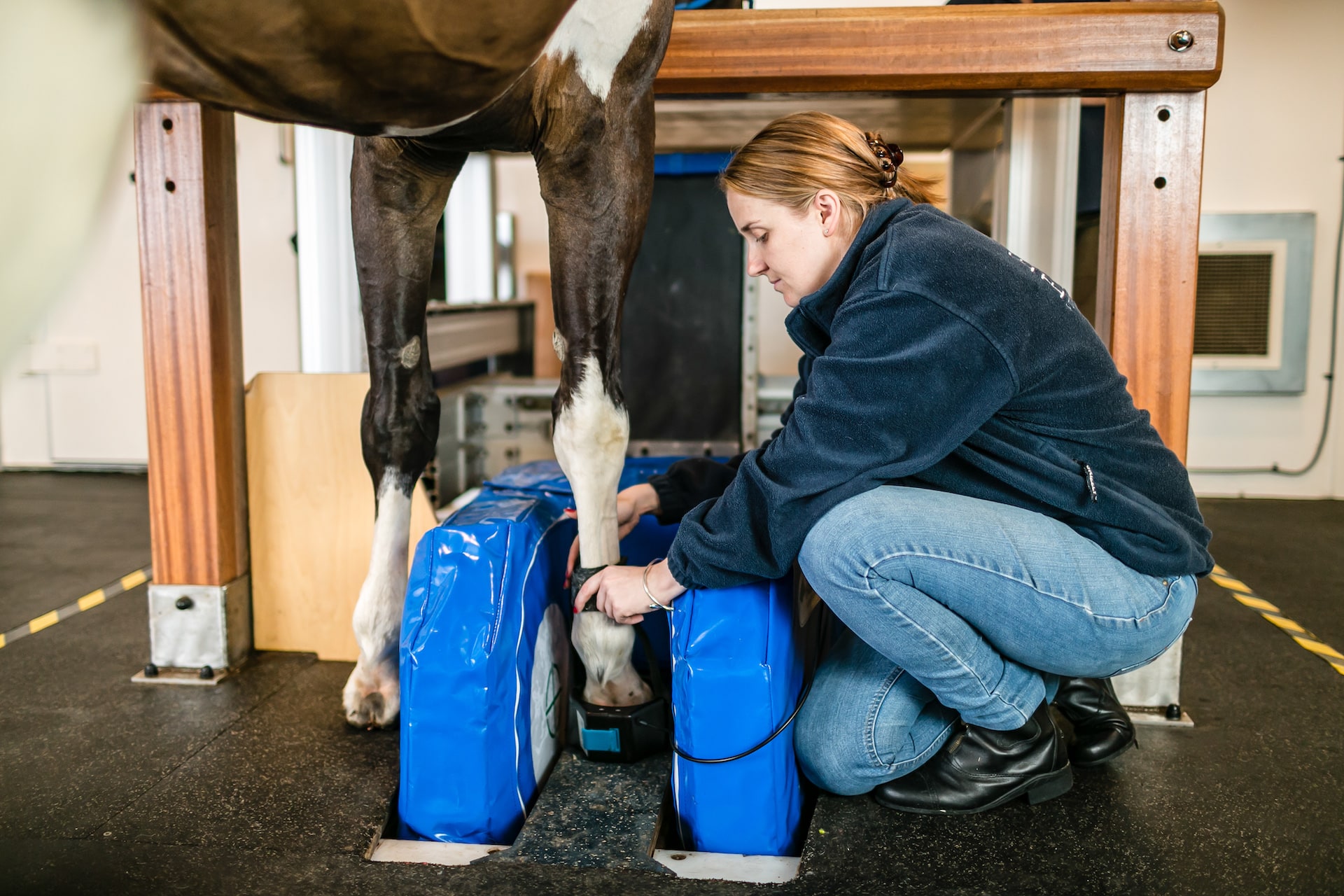
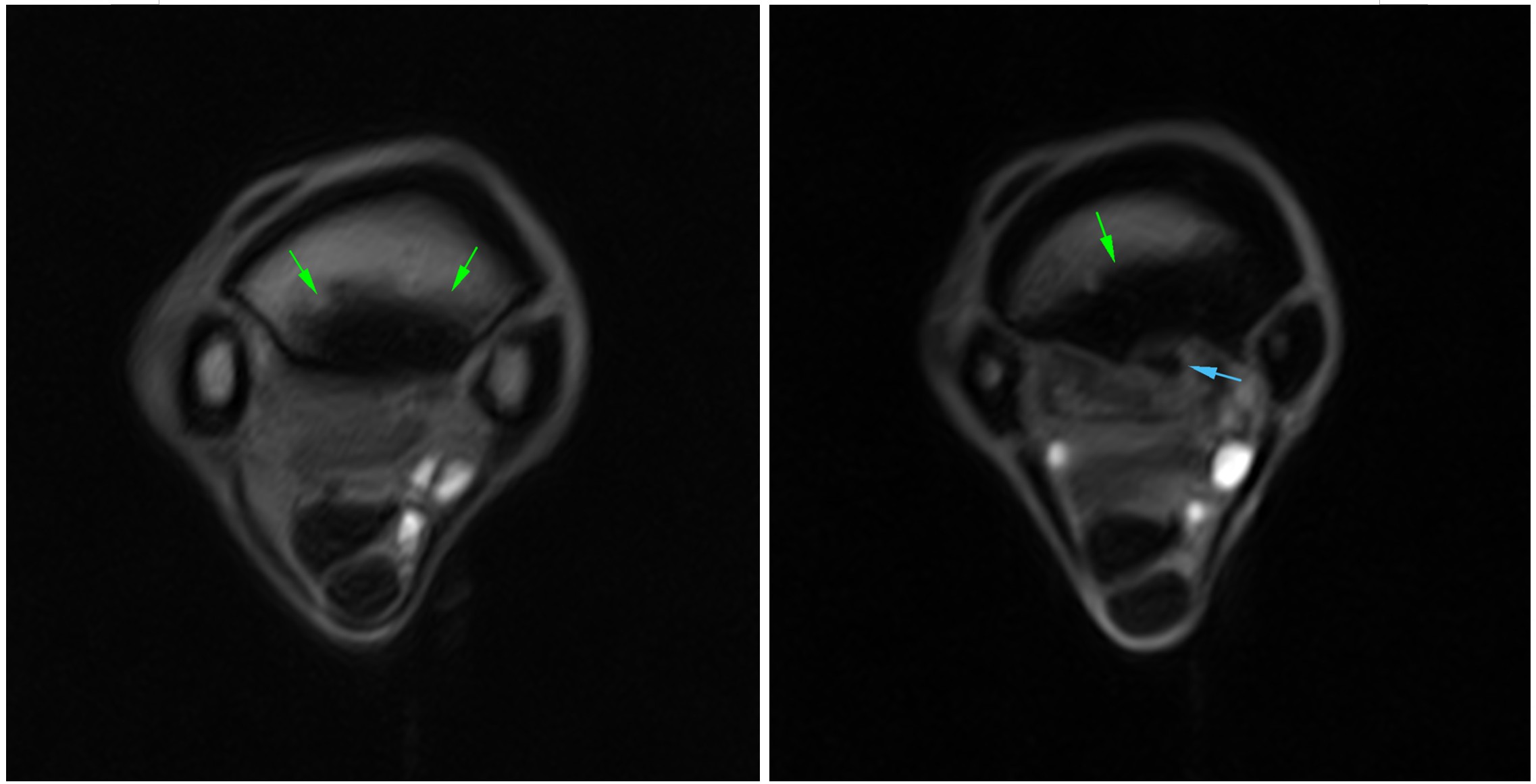
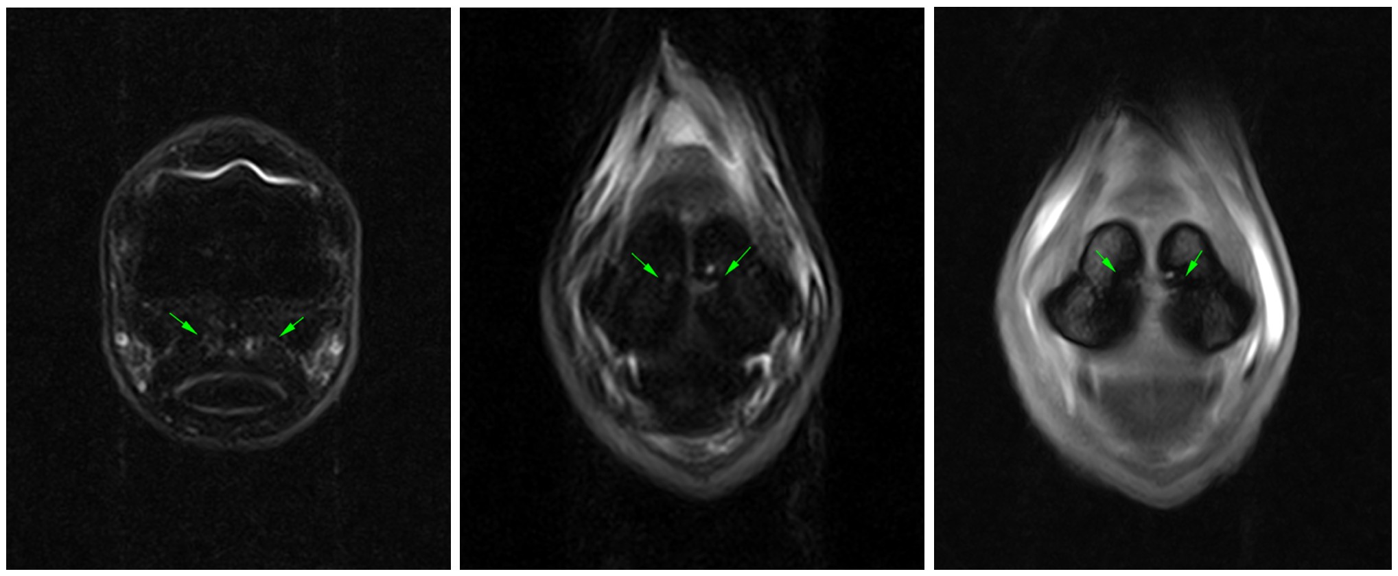
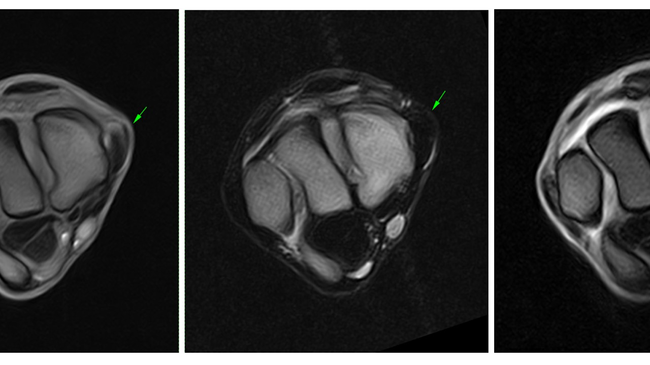
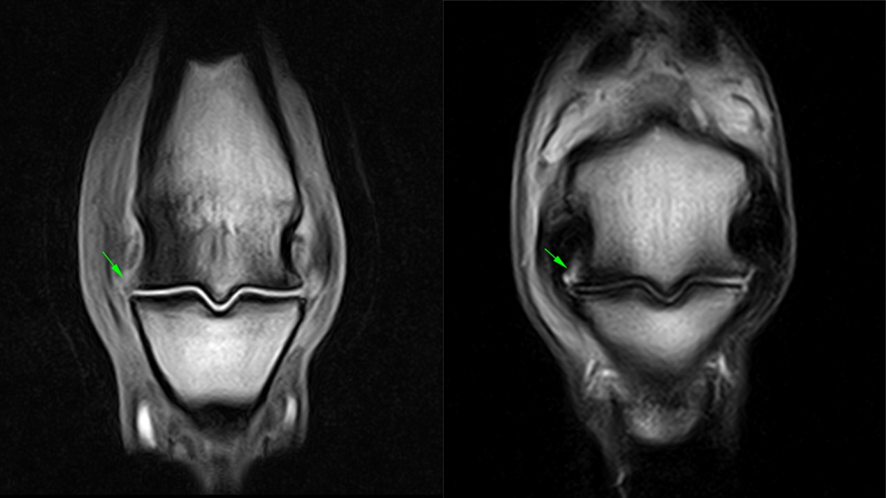
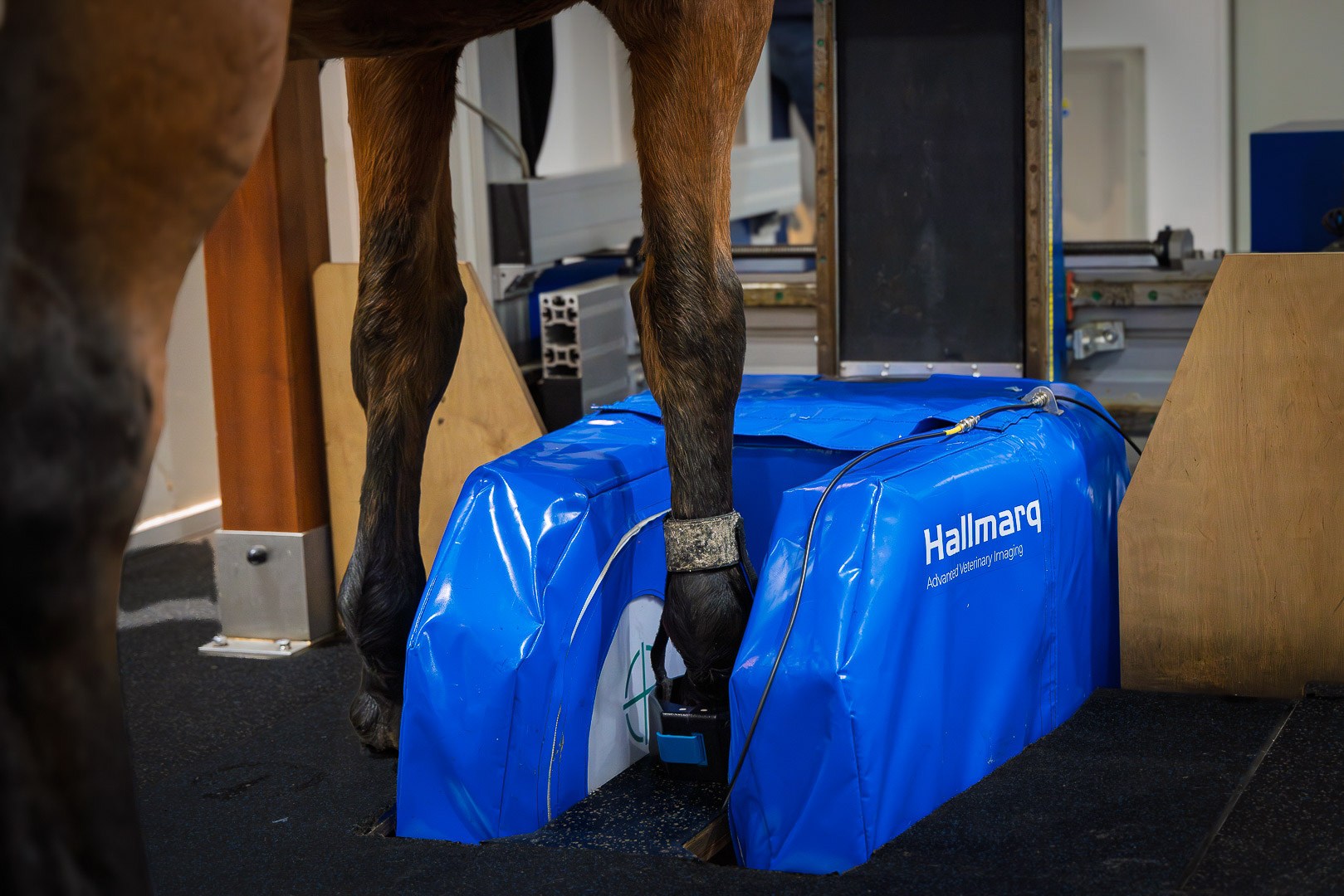
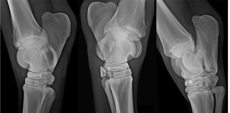
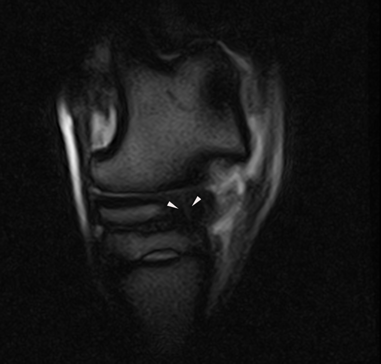
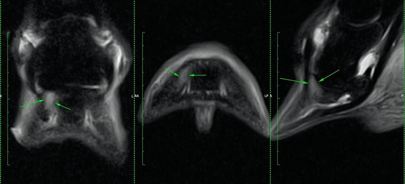
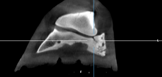
Skip to the content

Looks like you’re on our UK site! Would you like to switch US site?

Do not ask again
Switch to US

Welcome to the next generation!

Introducing the World’s First Veterinary-Specific Zero-Helium (ZHM) Small Animal 1.5T MRI

Find out more
Header image

Region

  • English (UK)
    • English (USA)
Community
Hallmarq Veterinary Imaging
Innovative diagnostic imaging solutions
  • Products & Services
    • Standing Equine MRI
    • Standing Equine Leg CT
    • Small Animal MRI
  • Solutions
    • Q-Care
    • MRI – for Equine Vets
    • MRI – for Horse Owners
    • CT – for Equine Vets
    • CT – for Horse Owners
    • For Small Animal Vets
    • For Pet Owners
  • News
  • Resources
    • Case studies
    • Webinars
    • Download centre
  • About
    • About us
    • Our team
    • Find a Hallmarq Site
    • Environmental, Social and Governance (ESG)
  • Contact
Search
Menu

Case Studies

Explore current trends and benefit from our users’ clinical results
Close search
Close
  • Products & Services
    Show sub menu
    • Standing Equine MRI
    • Standing Equine Leg CT
    • Small Animal MRI
  • Solutions
    Show sub menu
    • Q-Care
    • MRI – for Equine Vets
    • MRI – for Horse Owners
    • CT – for Equine Vets
    • CT – for Horse Owners
    • For Small Animal Vets
    • For Pet Owners
  • News
  • Resources
    Show sub menu
    • Case studies
    • Webinars
    • Download centre
  • About
    Show sub menu
    • About us
    • Our team
    • Find a Hallmarq Site
    • Environmental, Social and Governance (ESG)
  • Contact

Region

  • English (UK)
    • English (USA)
Community
  • All case studies
  • Equine MRI
  • Small Animal MRI
  • Equine CT

Hyperextension of the right front metacarpophalangeal joint

A vet with a horse in an MRI scanning room

A 9-year-old broodmare with a persistently draining solar abscess from her right hind foot

A left hind limb lameness resulting from a thrombosed digital artery

Thoroughbred mare with an acute exacerbation of an intermittent low-grade right hindlimb lameness

MRI operator, managing horse in Equine MRI scanner

Bilateral chondral lesions of the distal interphalangeal joint in a warmblood

Horse in Equine MRI scanner

Investigation of an osteolytic lesion on the medial border of the distal phalanx

A horse being scanned by the equine MRI machine

10-year-old Irish Sports horse with an inflammatory bone lesion in the pastern

Home > Case Studies > Case Studies > Equine MRI

Keep up to date

Improving the lives of animals and the people who care for them.

Email

email hidden; JavaScript is required

Follow Us

  • Facebook
  • LinkedIn
  • Twitter
  • Instagram

Product & Services

  • Standing Equine MRI
  • Standing Equine Leg CT
  • Small Animal MRI

Solutions

  • Q-Care
  • MRI – for Equine Vets
  • MRI – for Horse Owners
  • CT – for Equine Vets
  • CT – for Horse Owners
  • For Small Animal Vets
  • For Pet Owners

About

  • About us
  • Our team
  • Find a Hallmarq Site
  • Environmental, Social and Governance (ESG)

Copyright © Hallmarq Veterinary Imaging 2023

Registered in England and Wales Company No. 4061791

  • Privacy Policy
  • Cookie Policy
  • Terms of Use
  • Sitemap

Web design Surrey

Please select your Community

Don’t worry, you can change your selection at any time.

Equine

All the information and content covering equines.

Join Community

Small Animal

All the information and content covering small animals.

Join Community