

Historically, standing MRI has most commonly been used for investigation of foot lameness. However, in the case of this patient, the sequences obtained with MRI allowed detailed evaluation of soft tissue structures at the more proximal level of the limb. A diagnosis of collateral ligament desmopathy of the carpus was made. Let’s take a look…
History
An 8-year-old Thoroughbred gelding presented with acute right forelimb lameness after briefly escaping from the yard; a traumatic event could not be excluded. Perineural analgesia of the lateral palmar nerve was negative. However, lameness improved following median and ulnar nerve block and intra-articular anaesthesia of the radiocarpal and middle carpal joints.
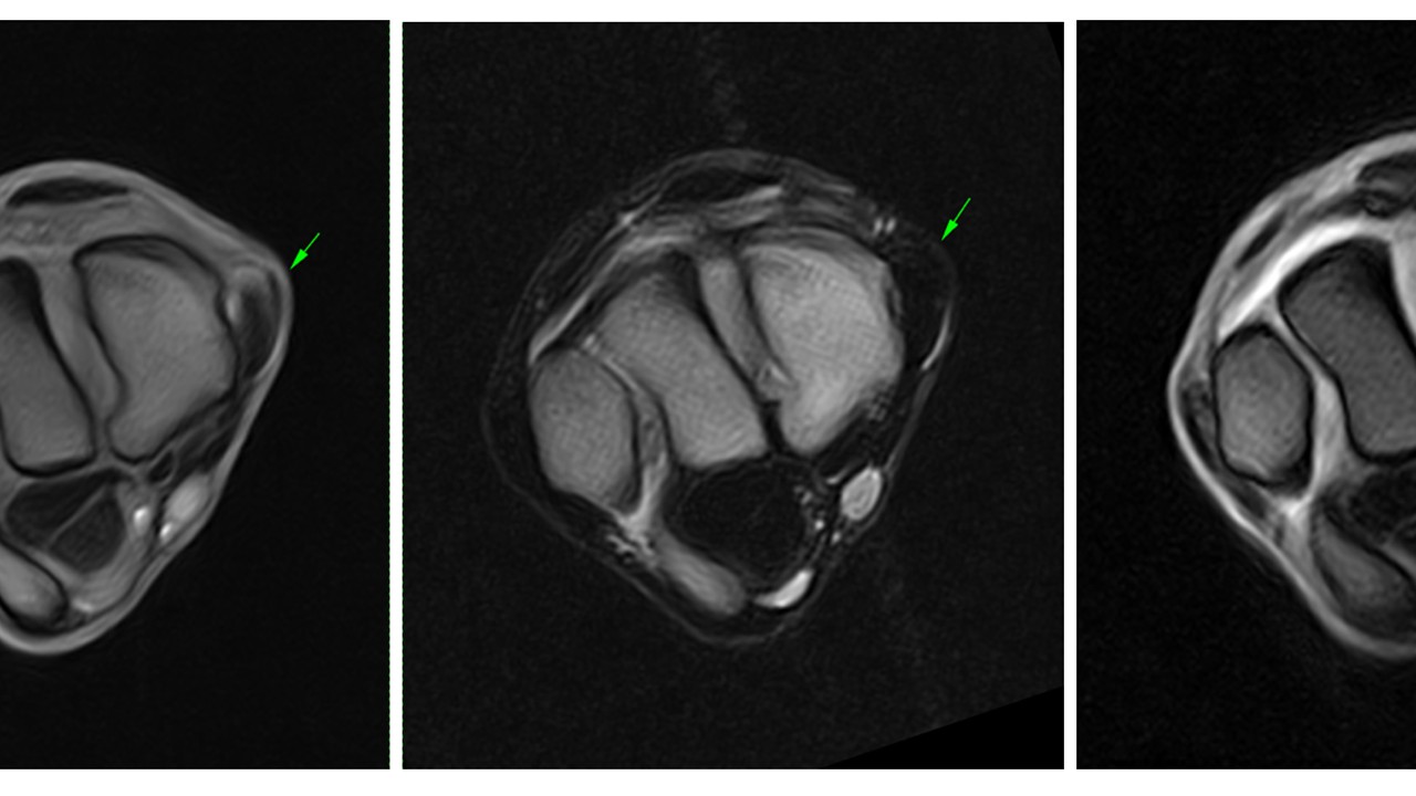
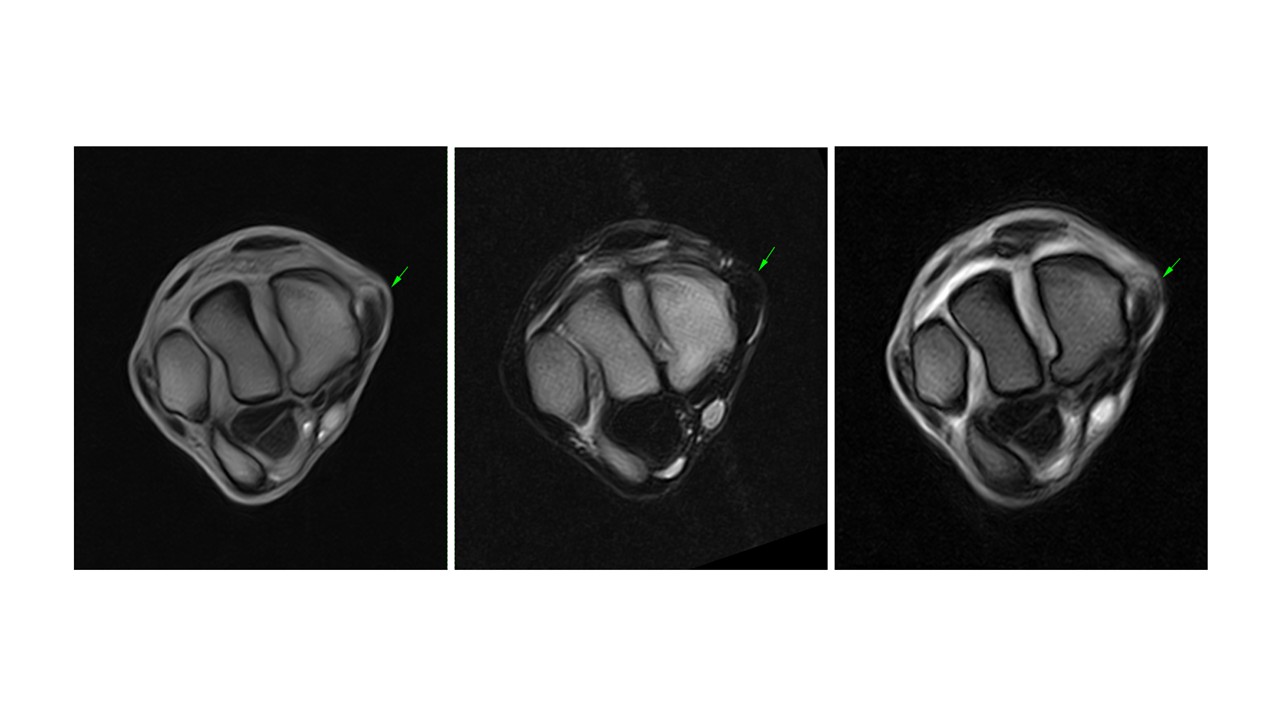
MRI findings
The horse underwent standing equine MRI of the right carpus. This revealed thickening of the proximal third of the medial collateral ligament of the carpus. The dorsal margins of the ligament were irregular, with moderate increased signal intensity on the dorsal aspect on T1W and T2*W GRE sequences and mild increased signal intensity on T2W FSE sequences. The periligamentous soft tissues were minimally thickened. The distal portion of the ligament appeared normal.
There was no evidence of joint effusion or subchondral or trabecular bone abnormalities. The included portion of the suspensory ligament was normal.
Conclusion
The findings were consistent with desmopathy of the medial collateral ligament of the carpus involving its proximal third. MRI identified an unusual ligamentous lesion that was considered the most likely cause of the lameness. In addition, it excluded other significant osseous or soft tissue injury within the carpus. Although standing MRI is most commonly used for investigation of foot lameness, the sequences obtained allowed detailed evaluation of soft tissue structures at this more proximal level of the limb.
A traumatic aetiology was suspected based on the clinical history.
With thanks to Dr Marianna Biggi and Dr Federica Cantatore, Pool House Equine Hospital, UK for sharing this case with us.


