

History
The horse was referred to the clinic for investigation of right forelimb lameness. The mare had undergone MRI examination of the carpus four months earlier, which revealed mild enlargement of the accessory ligament of the superficial digital flexor tendon (SDFT) and moderate distention of the carpal sheath.
MRI findings
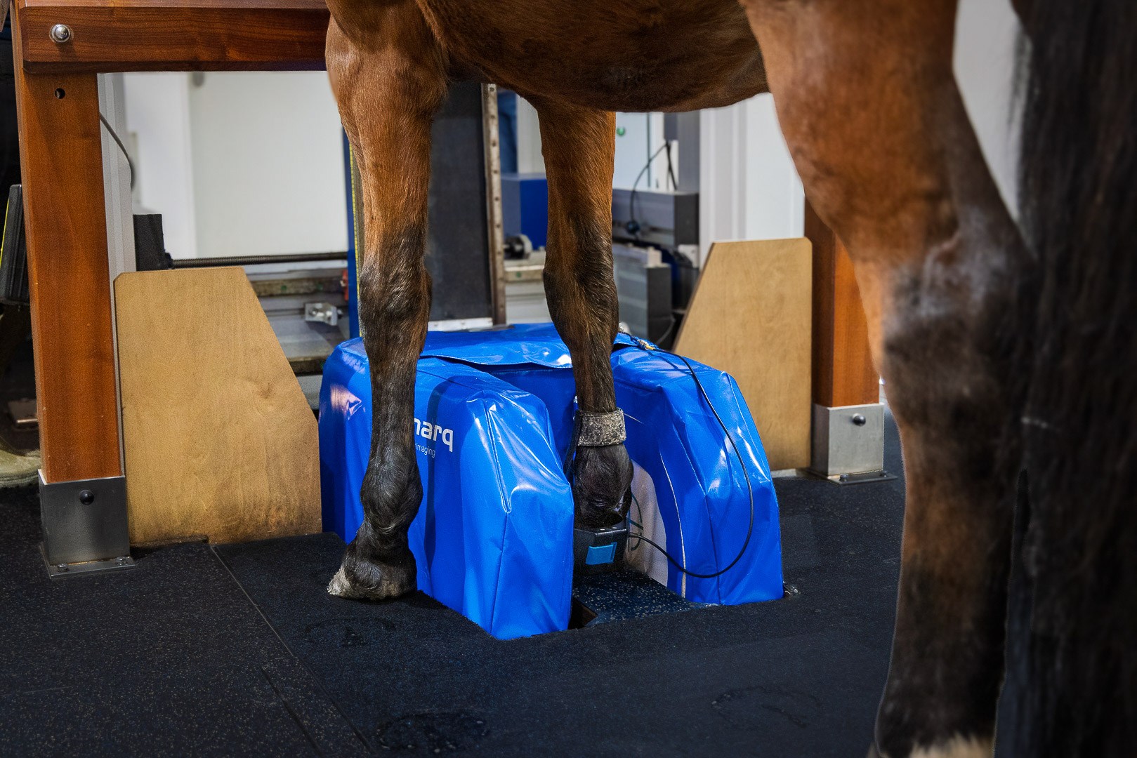
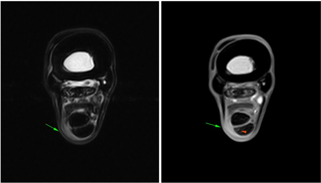
The horse underwent standing equine MRI of the right proximal metacarpus and carpus. Beginning approximately 40 mm distal to the carpometacarpal joint and extending for approximately 40 mm proximodistally along the lateral margin of the flexor tendons, there was thickened peritendinous soft tissue material measuring approximately 5 × 18 mm. This material was hyperintense on T2*W sequences and moderately hyperintense on T2W sequences. Adjacent to this region, the lateral margin of the SDFT was mildly irregular and mildly heterogeneously hyperintense on all sequences, consistent with mild tendinitis.
The carpus was similar in appearance to the previous MR images. Within the middle carpal joint, the distal subchondral bone surface of the radial carpal bone and the opposing surface of the third carpal bone were moderately thickened and hypointense on all sequences. Small to moderately sized periarticular osteophytes were present along the medial margin of the middle carpal joint, and the carpal joints were minimally distended. There was minimal distension of the carpal sheath.
Conclusion
The MRI findings were consistent with peritendinous thickened soft tissue material, most likely fibrosis, along the lateral margin of the flexor tendons within the proximal third of the metatarsus, with mild associated tendinitis of the SDFT. Distension of the carpal sheath had reduced compared with the previous examination. The clinical relevance of these findings should be interpreted in conjunction with the clinical examination.
With thanks to Suzy Hall, Liphook Equine Hospital, UK for sharing this case with us.


