


History
A 9-year-old Connemara horse was presented with a 2–3 month history of right forelimb lameness, most recently graded 2–3/10. The horse had a positive lateral palmar nerve block and had previously been treated for proximal suspensory ligament desmopathy. Ultrasonographic examination revealed enlargement of the superficial digital flexor tendon (SDFT) but the fibre pattern was not consistent with an acute injury there was no pain on palpation of the region.
MRI findings
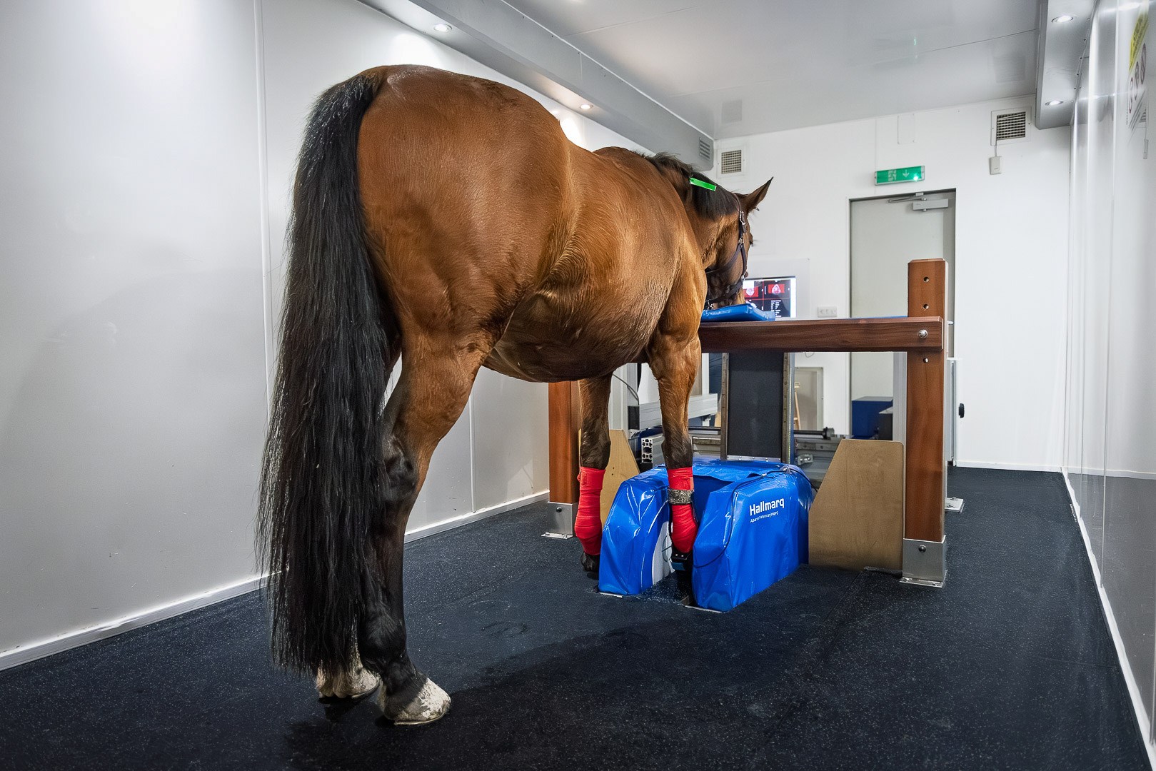
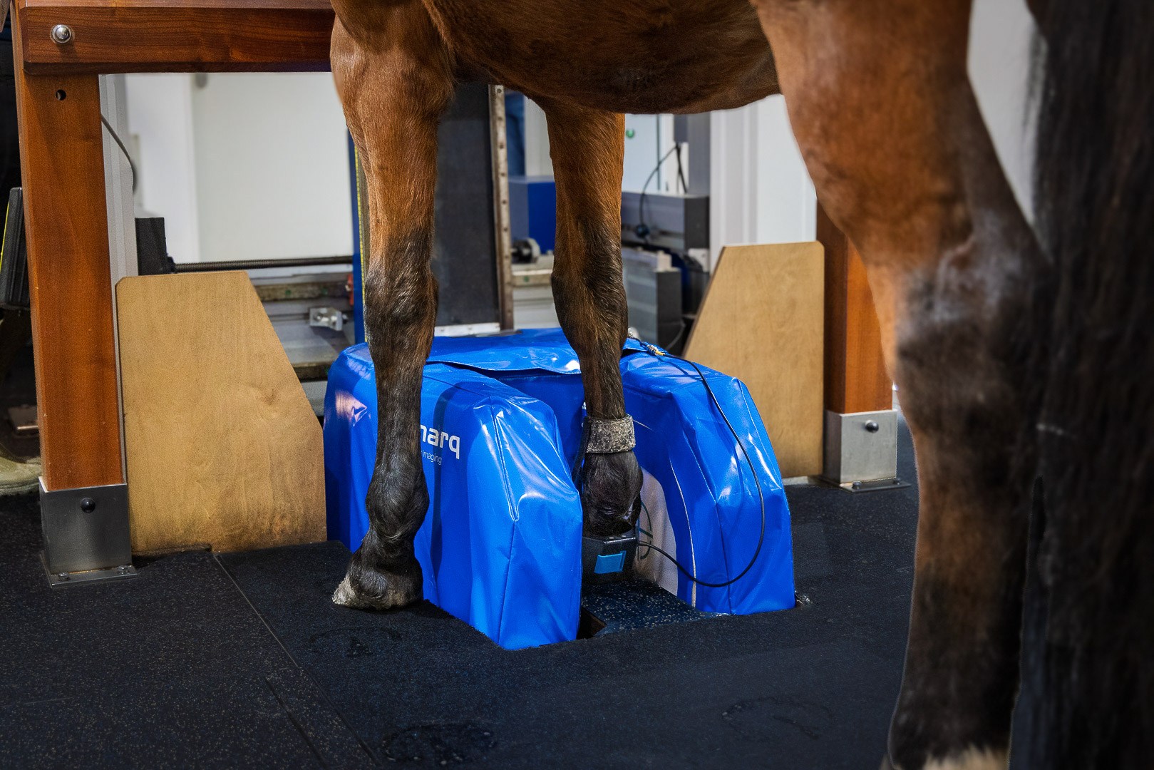
The horse underwent standing equine MRI of the right metacarpal and carpal regions. This revealed mild enlargement of the lateral aspect of the SDFT in the proximal third of the metacarpal region.
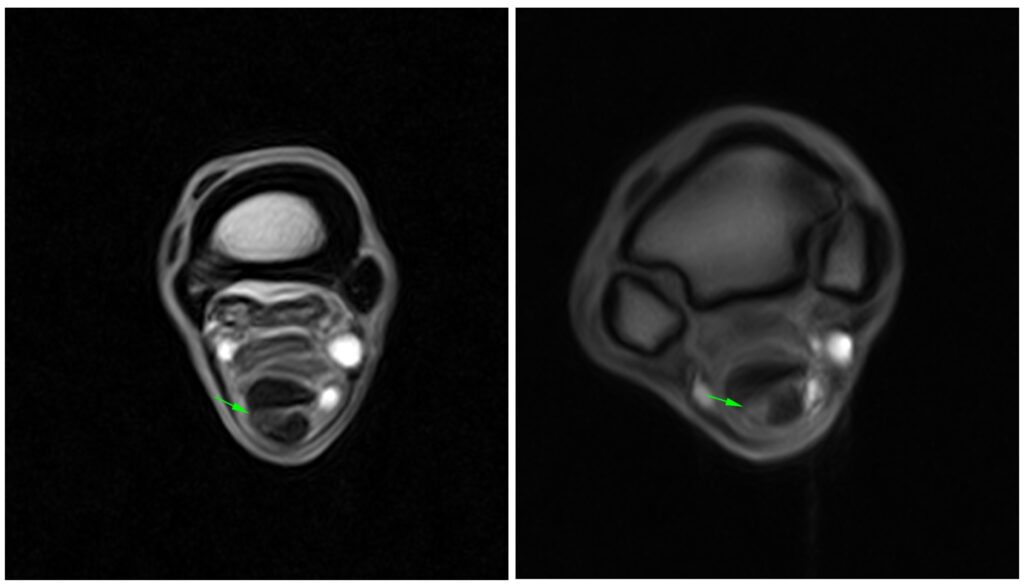
The borders of the SDFT were poorly defined with mild ill-defined increased T1W signal within the lateral half of the tendon and ill-defined increased T2W signal intensity in the lateral aspect of the proximal SDFT.
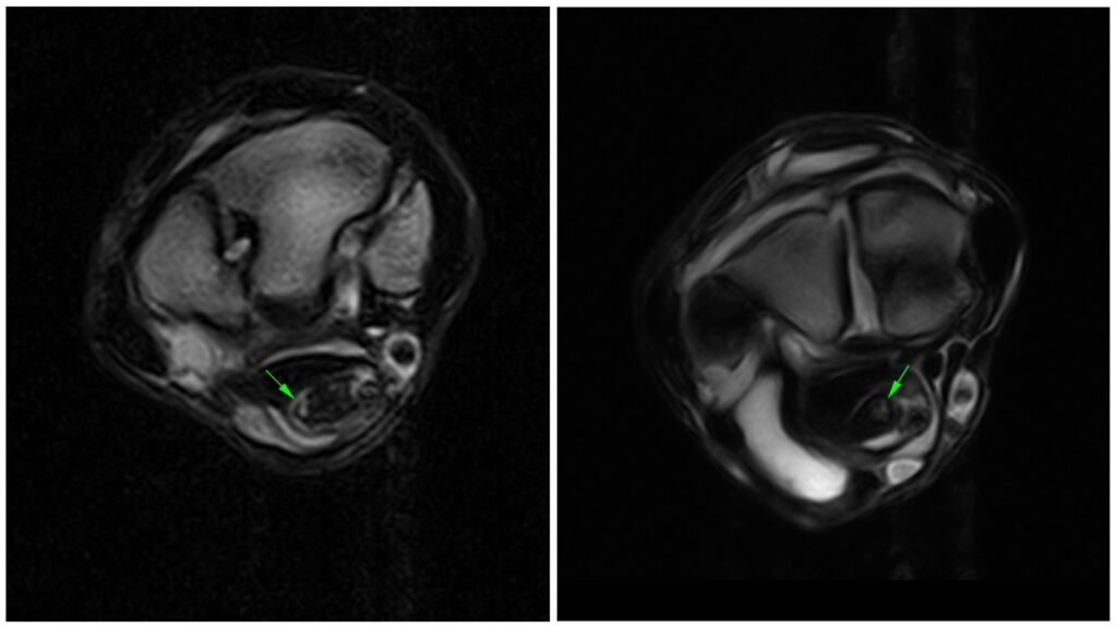
These changes extended proximally to the level of the distal row of the carpal bones, where there was mild diffuse enlargement of the tendon, mild patchy increased T1W signal intensity in the medial half of the SDFT, mild patchy increased T2W signal within the tendon and mild increased lateral peripheral STIR signal.
The poor lateral border definition and patchy increased T1W signal continued proximally to the proximal row of carpal bones with very mildly increased peripheral T2W signal. At the proximal aspect of the proximal row of carpal bones there was patchy increased T1W and T2W signal in the dorsomedial aspect of the SDFT extending to the level of the distal radius. There was moderate fluid distention of the carpal sheath.
Conclusion
In this study of proximal SDF tendinopathy, the findings were consistent with chronic tendinopathy of the superficial digital flexor tendon at the proximal third of the metacarpal region. However, at the proximal extent of the proximal third of the metacarpal region and extending adjacent to the carpus, the imaging findings suggested replacement of the normal low signal intensity collagen by fluid, such as oedema, haemorrhage, increased vascularity or necrosis. The proximal SDF tendinopathy (level 0) was likely to be relevant to the lameness in the horse.
The different MRI sequences aided lesion characterisation for the soft tissue injury, as the more distal component of the lesion demonstrated predominantly increased signal intensity on T1 weighted sequences consistent with chronic tendinopathy, whereas the more proximal region also showed signal alterations on T2 FSE and STIR sequences, indicating increased fluid signal within the tendon.
With thanks to Dr Ceri Sherlock and team at Bell Equine veterinary Clinic for sharing this case with us.