

We review a case of extensive mineralization of the collateral sesamoidean ligament kindly provided by Valley Equine Hospital.
The study looks at a large breed horse who was referred for imaging with Hallmarq’s Vision CT due to persistent left forelimb lameness that had not resolved despite various rehabilitation and treatments. The horse exhibited lameness at the walk and had a positive response to a high palmar digital nerve block.
CT findings
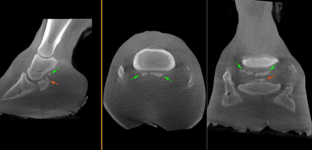
The main findings were marked, multifocal mineralization of the collateral sesamoidean (navicular suspensory) ligament, with marked enthesophytes and an avulsion fracture of the proximal aspect of the navicular bone. There was a very small focal region of calcification palmarly to the navicular bone, suspected to be within the deep digital flexor tendon. There was additional marked ossification of both collateral cartilages.
The outcome
A palmar digital neurectomy was elected as the treatment of choice, given the horse’s 80–90% improvement following the nerve block and the lack of success with previous conservative treatments. The surgery was performed only for the horse to return to a life as a pasture horse and not as a ridden horse given the extent of the damage and the risks being higher than usual. Additionally, the horse was shod with remedial farriery, appropriate for managing navicular changes.
Conclusion
In this particular case of extensive mineralization of the collateral sesamoidean ligament, the horse’s large foot size made imaging with standing MRI unsuitable. As the foot would not fit in the scanner, any resulting images would likely be non-diagnostic. Trimming of the foot was necessary to acquire the CT images. The foot is a complex anatomical structure where radiographs often prove inadequate due to superimposition of structures. Even with additional non-standard views, pathology is frequently underestimated.
Vision CT was considered the ideal imaging modality in this case, as it provides superior visualization of mineralization compared to MRI and enables 3D reconstruction compared to radiographs, offering a comprehensive diagnostic advantage. Despite some compromise in image quality due to the size of the foot, a definitive diagnosis was successfully reached.
With thanks to Dr Tom McParland BVetMed DiplACVS (LA) MRCVS and Melissa Lockwood RVN, Valley Equine Hospital, UK, for sharing this case with us.