

Technology
How it works
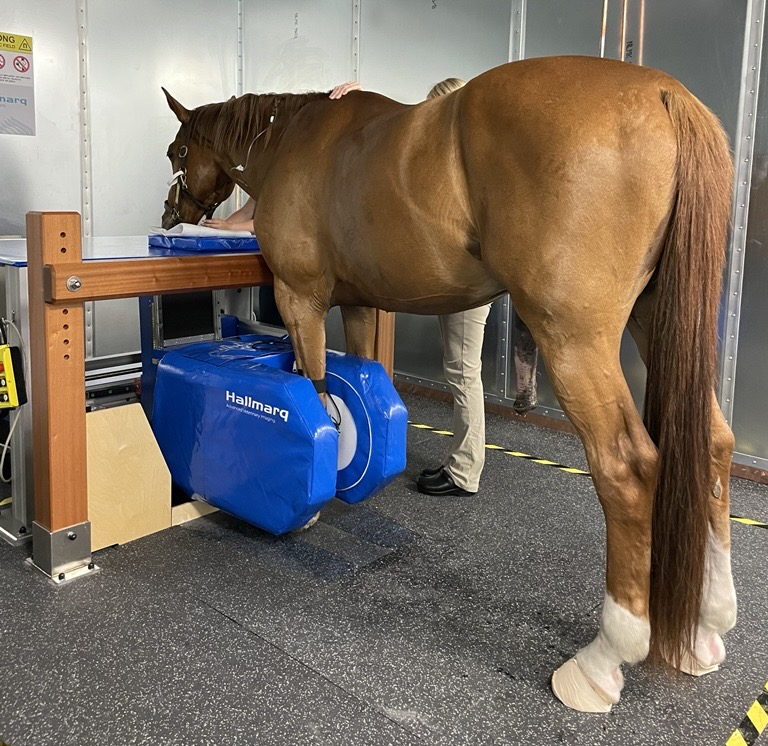
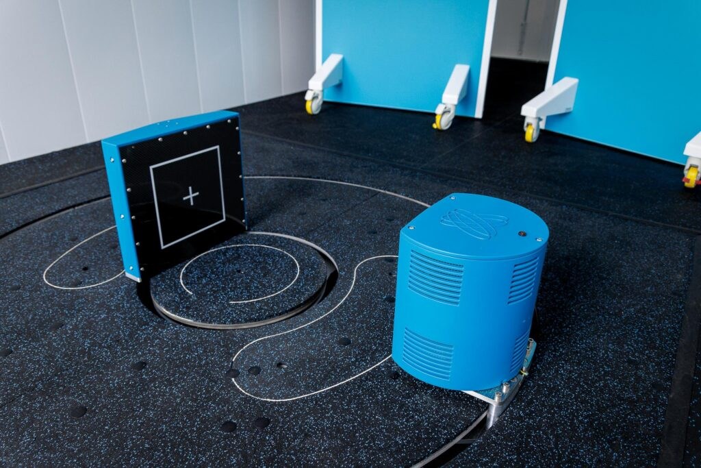
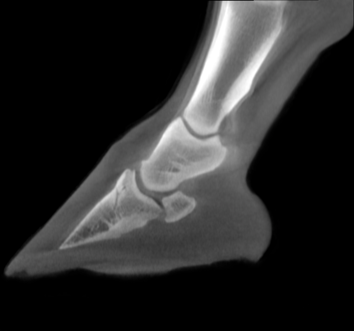
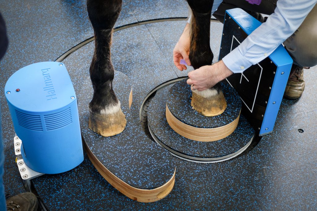
Designed to image the distal limb of the standing, sedated horse, Vision CT captures 3D image sets in just minutes. With our walk-in-scan-walk-out design, the horse is unrestrained with easy entry and exit for safe, effective, and affordable advanced imaging
Details
Clinical uses
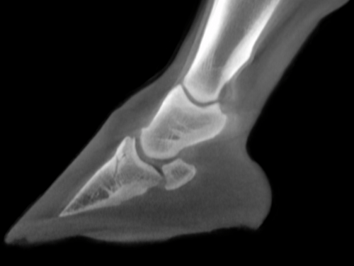
Surgical planning
Advanced pre-operative imaging to help determine the precise nature and location of pathology
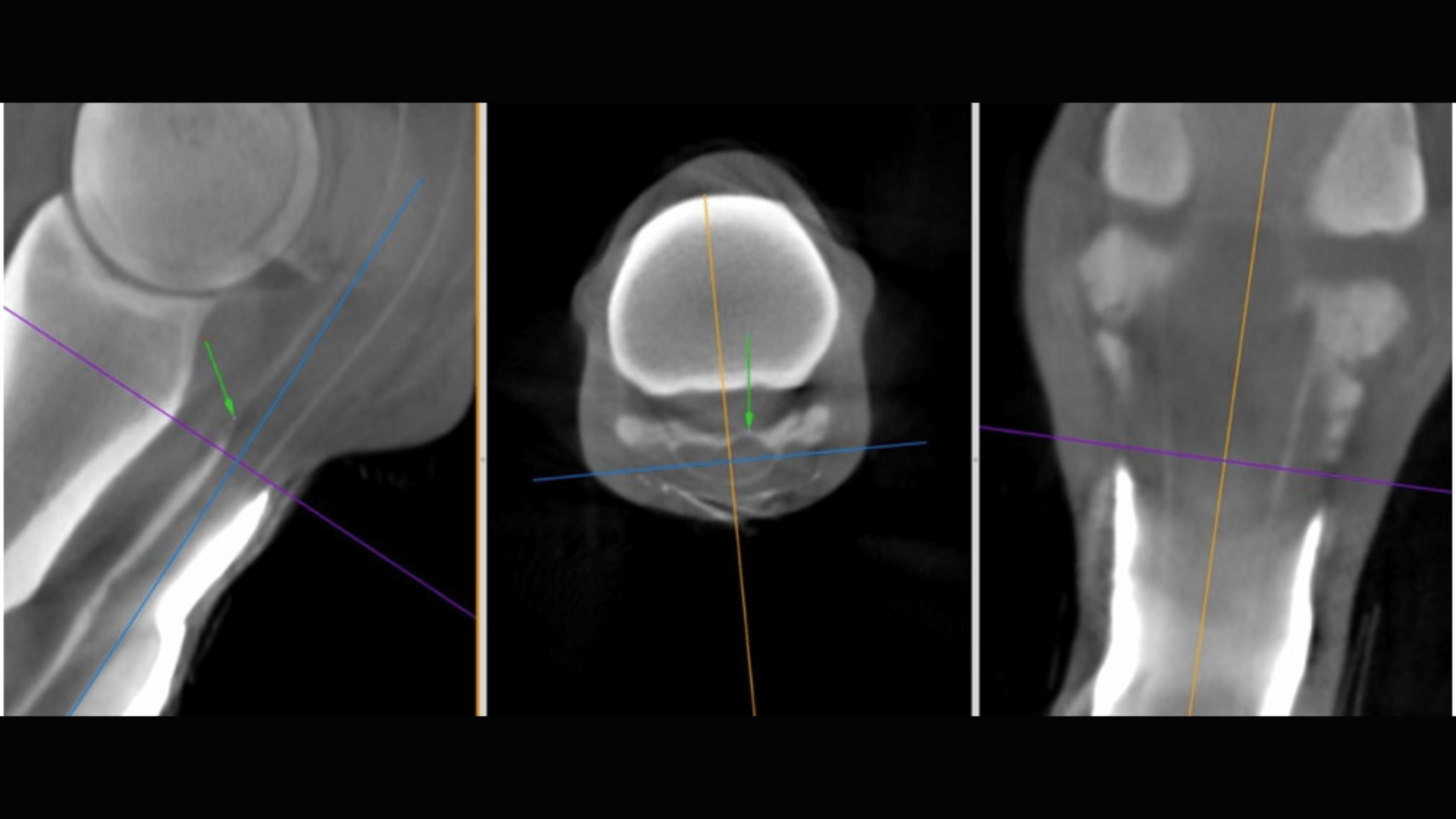
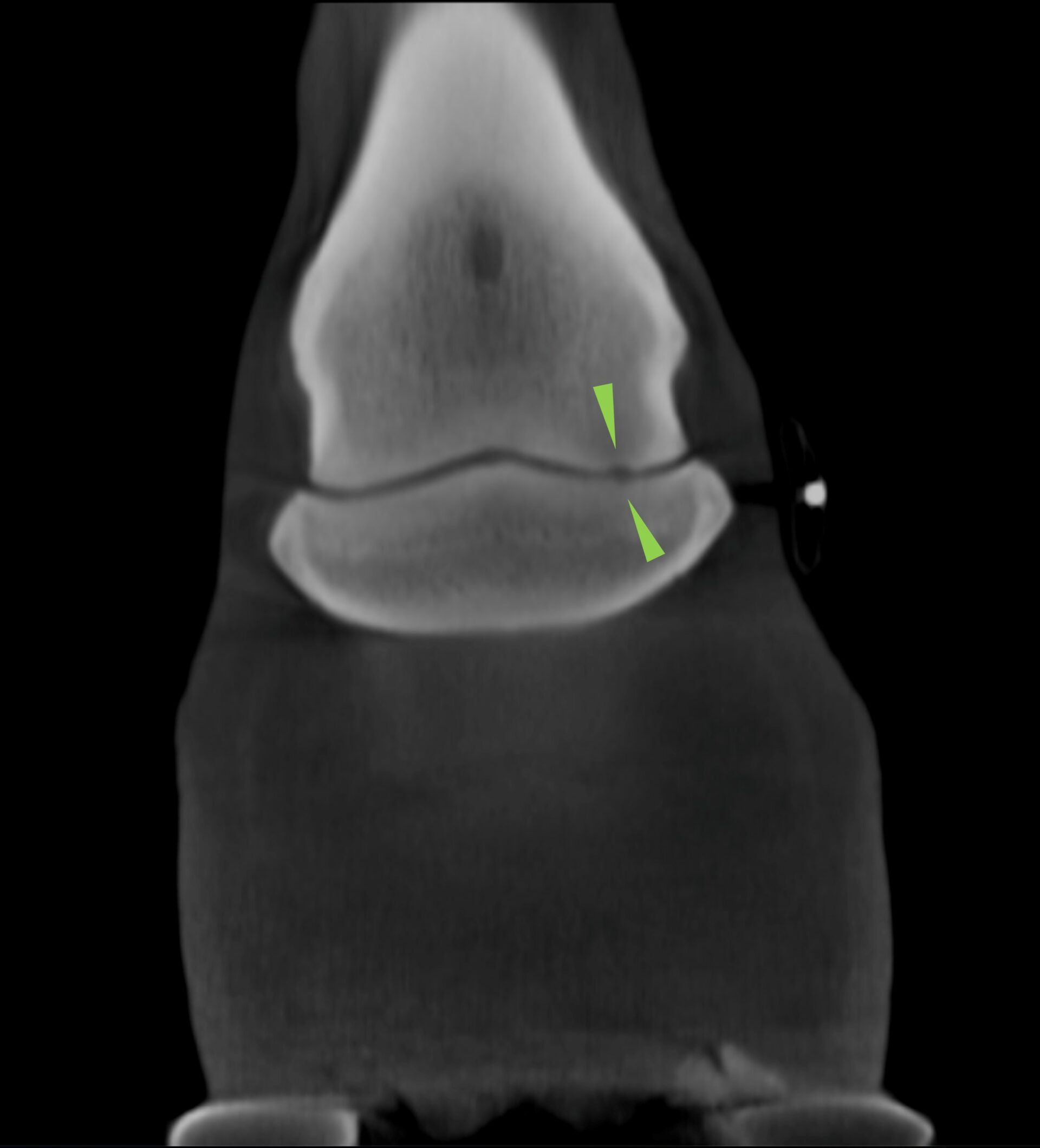
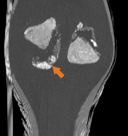
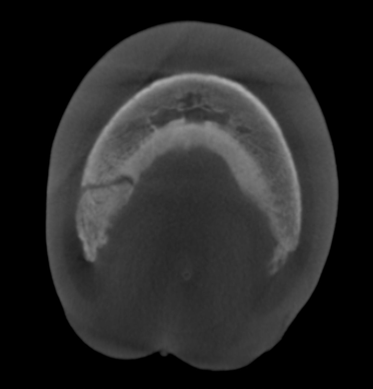
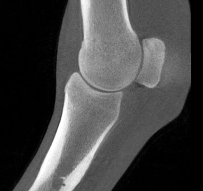
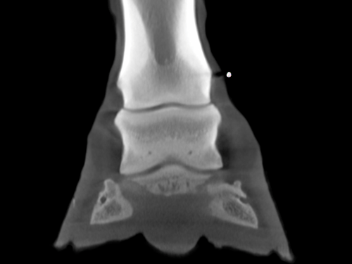
Bony tissue differentiation
Detect non displaced fractures, small osteophytic lesions and evaluate cortical and sub-chondral bone
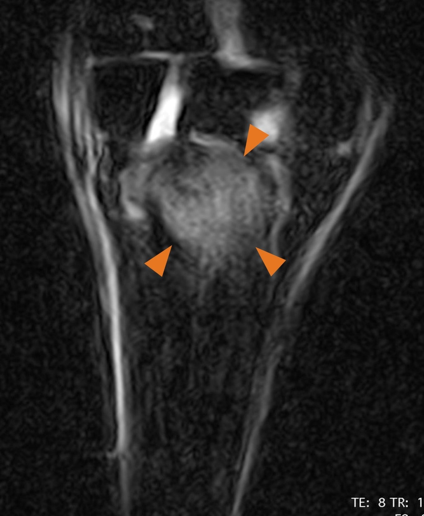
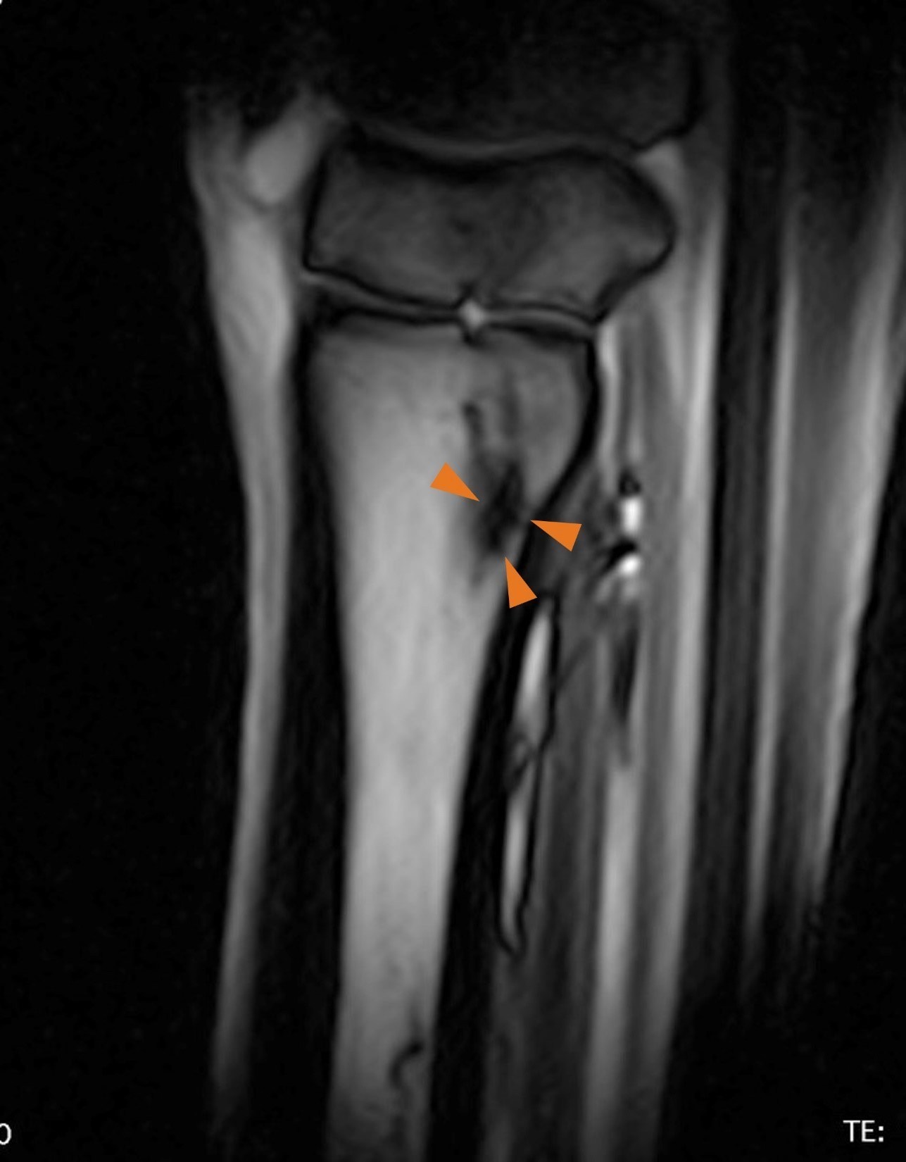
The complete picture
Obtain complementary clinical information when combined with Hallmarq’s unique Standing Equine MRI
Why CT?
Features & benefits
Safe for horse & handler
The walk-in, walk-out floor-level design allows for easy entry and exit. With no general anaesthesia required and low-dose radiation, both handler and horse are safe throughout the procedure.
Effective imaging
High spatial resolution detects small changes in the distal limb without anatomical overlap or distortion. Images in standard DICOM format allow flexible viewing from any angle.
Affordable
With minimal installation and operating costs, there’s no need for a custom-built room. Monthly payment plans make Vision CT profitable with as few as two cases per week.
Fast acquisition
The system captures high-quality 3D image sets in just minutes. Combined with our motion correction software, this enables faster and more accurate diagnosis than traditional radiographs.
Space efficient
The compact footprint requires no specialised room, making installation easy even in smaller clinics. Offer advanced imaging without major renovations or disruptions.
Q-Care support
Responsive system support, for minimal downtime and maximum efficiency. Maintain smooth operations for increased workflow and profitability.
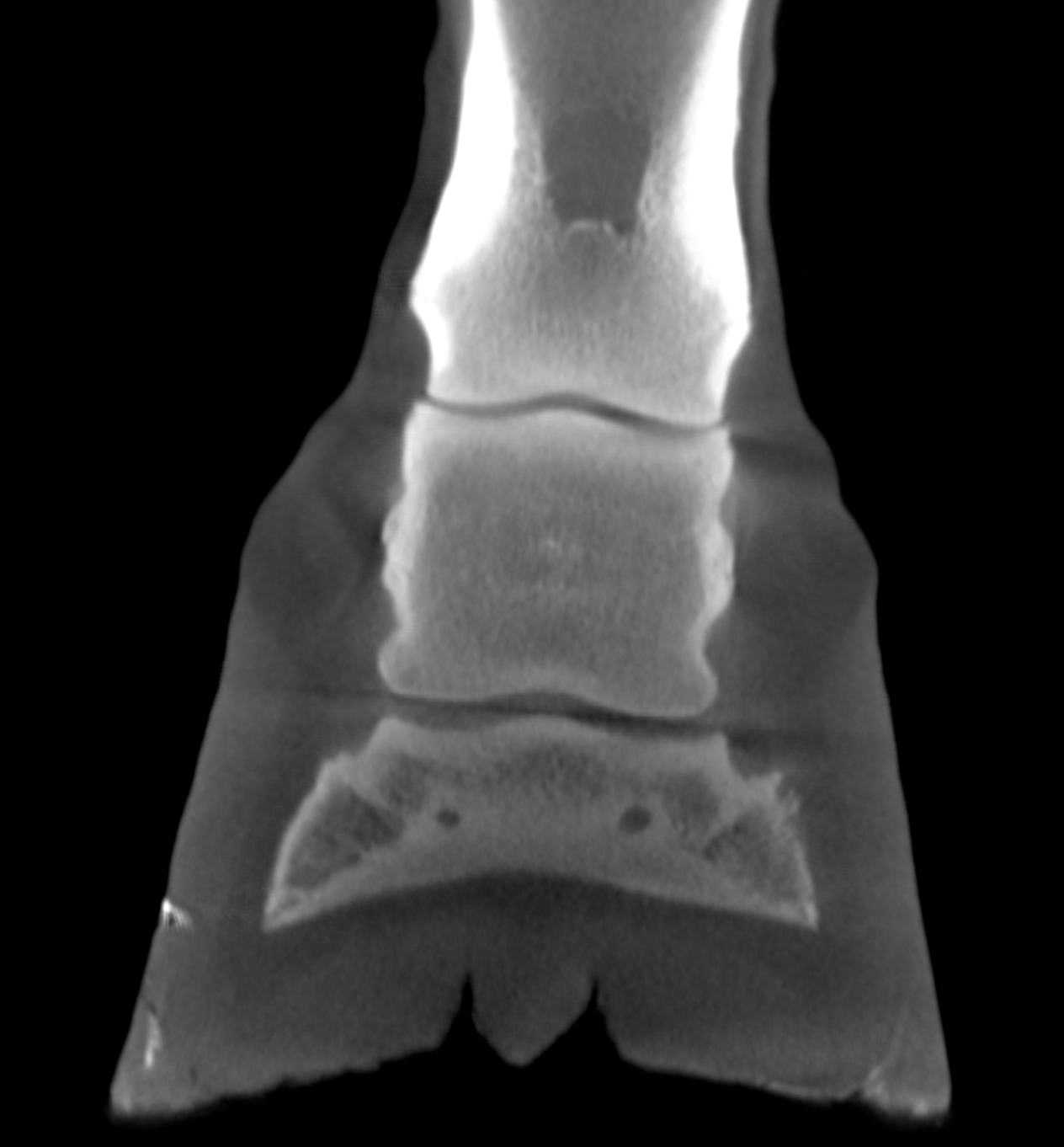
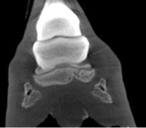
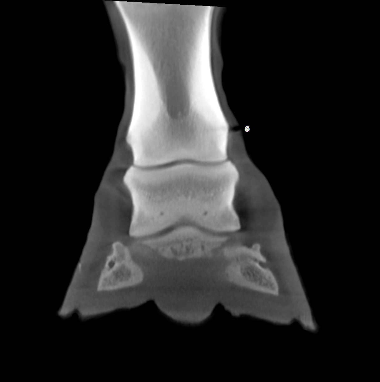
Detailed Imaging
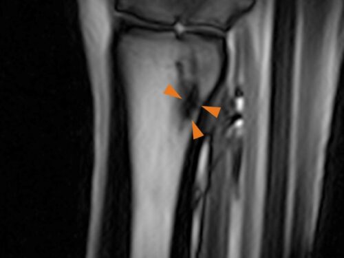
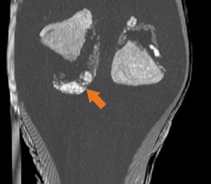
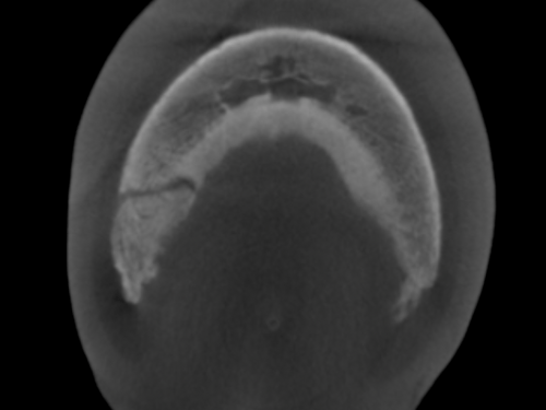
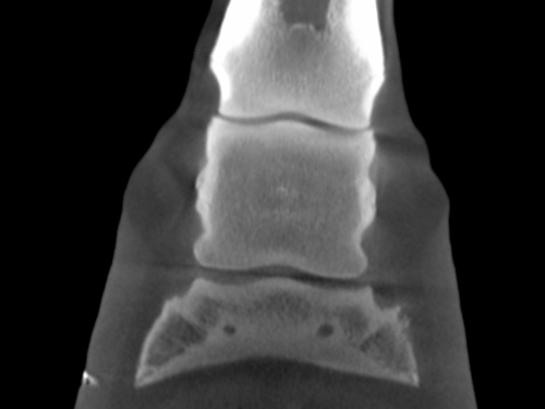
Clinical Gallery
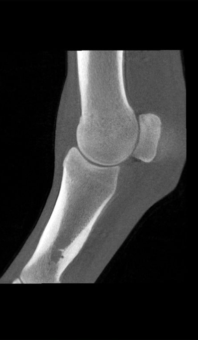
Specifically designed to image the distal limb of the standing, sedated horse, Vision CT captures images for a more accurate diagnosis.
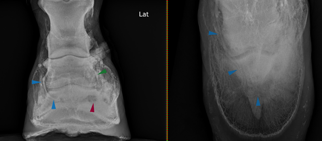
- Characterize foreign bodies in the hoof and assist with surgical planning
- Provide the answers in cases involving metallic objects
- Obtain a quantitative assessment of lesions and fracture lines
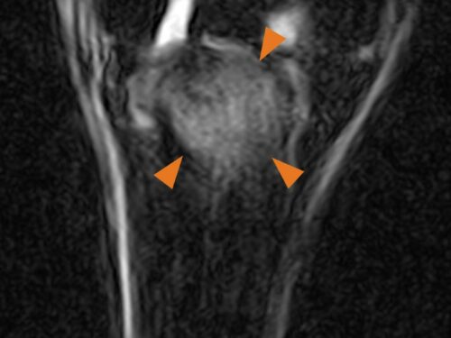
- Complement Standing Equine MRI
- Complement scintigraphic findings
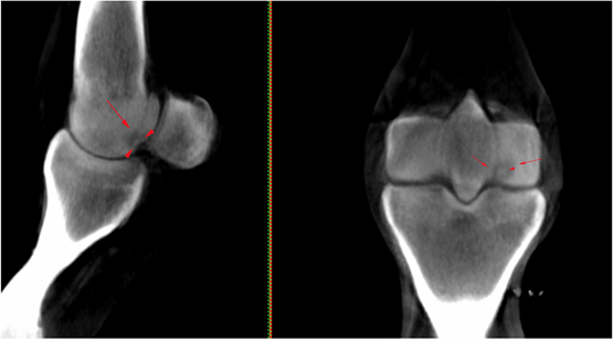
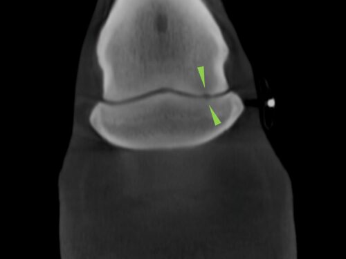
- Better evaluate and characterize irregularities on the articular surface
“The system was very straightforward to install and has a simple-to-use interface. Quick acquisition time and fine bone detail make surgical planning much easier.”
Jonathon Dixon BVetMed MVetMed Dipl.ECVDI MRCVS Rainbow Equine Hospital
“slCT really complemented what the MRI had told us about the content and signal intensity and, in this case, helped with surgical planning, providing useful information on the bone margin.”
Alison Fairburn Specialist in Diagnostic Imaging BVSc DipECVDI-LA MRCVS Bell Equine Veterinary Hospital
“We are really excited to put into practice the development so far to help facilitate the move into second phase clinical trials and offer our customers a world first.”
Jonathon Dixon BVetMed MVetMed DipECVDI MRCVS Rainbow Equine Hospital
“we are interested to see if the slCT can improve our ability to diagnose cartilage defects in coffin joints and fetlocks.”
Luis Rubio-Martinez DVM DVSc PhD DACVS DECVS DACVSMR MRCVS Sussex Equine Hospital
“Hallmarq’s CT has delivered 3D images of feet, pasterns and fetlocks in standing horses in a practical, quick and easy way.”
Ceri Sherlock BVetMed (Hons) MS MVetMed DipACVS-LA DipECVDI-LA DECVS MRVCS RCVS and European Specialist in Equine Surgery and Veterinary Diagnostic Imaging Bell Equine Veterinary Hospital
“The standing leg cone-beam CT system from Hallmarq provides a safe, and convenient diagnostic modality to help produce a definitive diagnosis and guide the best treatment plan for equine patients.”
Jonathon Dixon BVetMed MVetMed DipECVDI MRCVS Rainbow Equine Hospital
“The 3D information generated from Hallmarq’s standing CT has helped us with our diagnoses and provided more information than conventional imaging modalities in many cases already.”
Alison Fairburn Specialist in Diagnostic Imaging BVSc DipECVDI-LA MRCVS Bell Equine Veterinary Hospital
Safe
Keep horse and handler safe with our unique floor-level system for easy walk-in and walk-out access. With no need for general anaesthesia, the gently sedated horse simply walks into the system for easy image acquisition. With less radiation than traditional fan beam CT, the technician remains in the room – behind a shielded screen – throughout the process. The scan can be paused at any time for a safe and easy exit if needed.
Effective
Looking for high-resolution, cross-sectional images for clearer differentiation of bony pathology? Bypass conventional imaging and step up to fast 3D imaging with cone beam technology. Distinguished by its sub-millimetre resolution and ability to perform detailed 3D reconstruction, Vision CT can provide a definitive diagnosis for improved treatment planning.

Affordable
Reduce your investment risk with our unique business model. With low installation costs, low running costs and no special power supply required, you can run a profitable CT service. With comprehensive support included, enjoy high case throughput with fast acquisition times. And, as an outpatient procedure with no need for general anaesthesia, keep costs low and the demand on your team to a minimum.
FAQs
Frequently asked questions
Yes, whilst the Hallmarq Standing Equine Leg CT is accessible to most practices, it is also possible to refer patients. The referral clinic will need to know the case history and any previous diagnostic results. After a CT scan, they will provide an interpretation and radiological report. Other options may also be available by arrangement:
- suggestions regarding treatment and prognosis
- an explanation to the client, in the appropriate language
- further case management or treatment
Find a system
Our systems are available at some of the world’s leading veterinary facilities around the world. To find a Hallmarq MRI or CT machine near you, click below.
Find a siteDetails
Specifications
In planning for the installation of your Vision CT system, the high-level technical specifications below should be taken into consideration.
| System Specification | |
|---|---|
| Generator | Cone Beam |
| 80kV/1.25mA | |
| Form Factor | Open |
| Equipment moves in concentric circles | |
| Speed | 360 degrees in one minute |
| FOV | 200mm per side (and 160mm high) cuboid |
| 160mm high | |
| Slice Thickness | 0.25mm |
| Scan Velocity | 1 rpm |
| Current Coverage | Distal limb |
| Motion Correction | Yes |
| GA | Not required |
| System Components | |
|---|---|
| Complete CT System | Source/detector/platform/footrest |
| Computer/keyboard/mouse/software/cabling | |
| Two operator shields | |
| User Manual | |
| CT Mainframe | 375.90 kg |
| 2000mm L x 2000mm W x 250mm H | |
| Side Extension Platform | 72.99 kg |
| 1500mm L x 600mm W | |
| Front Extension Platform | 1000mm L x 2000mm W |
| Ramp | 68.00 kg |
| 1000mm L x 1900mm W | |
| Ramp Side Extension | 20.00 kg |
| 750mm L x 600mm W | |
| Console Unit | 610mm L x 1000mm W x 900mm H |
| Radiation Screen | 60mm L x 1210mm W x 1990mm H |
| Electronics | |
|---|---|
| Powered from a regular single phase household standard supply: UK – 110-230V 4A max USA – 110V Europe – 220V | |
| Internet connection required with 500KB minimum band width |
| Software | |
|---|---|
| Modern clinical user interface | |
| Simple and easy to use | |
| Choice of multiple scan types | |
| PACS integration | |
| Built in back-up data | |
| Motion correction algorithm for improved images | |
| User-selectable reconstruction options |
Q-Care
Supporting your success
Providing high-quality images is just the beginning. With an unrivalled programme designed to add value to your practice, we’re here to help optimise your imaging service – from initial enquiry through to installation and beyond.