

DDFT Marginal Tear
In this study, kindly provided by Donnington Grove Equine Vets, we take a look at the case of an 11-year-old Warmblood gelding who was admitted for further diagnostic imaging of the right fore fetlock region. The horse became acutely lame on the right forelimb with concurrent swelling of the digital flexor tendon sheath. Ultrasonography of the proximal pastern identified possible changes within the oblique distal sesamoidean ligament.
“Contrast tenography of the digital flexor tendon sheath has improved diagnoses of manica flexoria tears but pre-operative diagnosis of marginal tears of the deep digital flexor tendon within the digital flexor tendon sheath can be difficult. This case uses dual modality (Vision CT and Standing Equine MRI) 3D-imaging to provide a pre-operative diagnosis of a marginal deep digital flexor tendon injury.”
Dr Sarah Taylor (BVM&S MSc PhD Cert ES(Orth) DipECVS DipECVSMR MRCVS)
Senior Lecturer in Equine Orthopaedics, The Royal (Dick) School of Veterinary Studies, The University of Edinburgh
MRI Findings
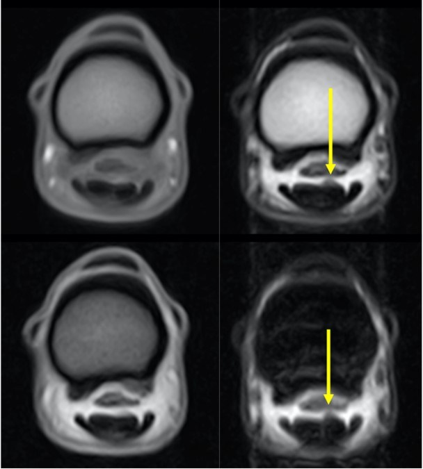
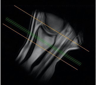
Figure 1: Transverse images of the proximal pastern; top left = T1 GRE FAST sequence, bottom left = T2* GRE FAST sequence, top right = T2 FSE FAST sequence and bottom right = STIR FSE FAST sequence. The yellow arrow highlights the abnormal dorsal curvature/enlargement of the lateral lobe of the deep digital flexor tendon with isointense signal (instead of the normal hypointense signal of the tendon) on T2 FSE and STIR sequences indicative of granuloma/torn tendon fibres with fluid signal presence. Figure 1(a) pilot shows the location of the slice.
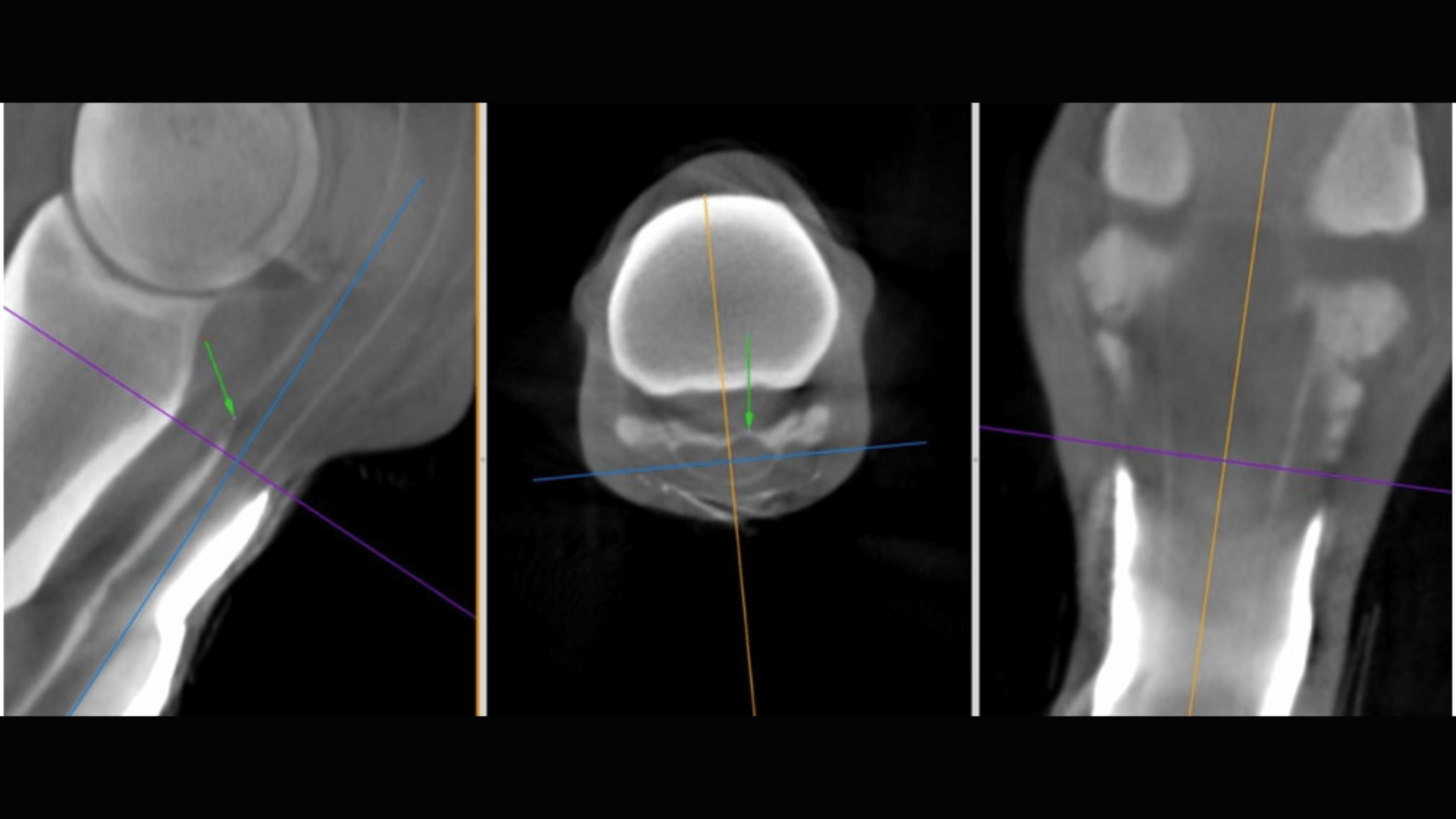
CT Findings
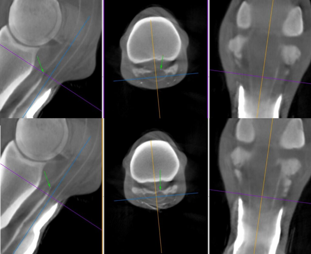
Figure 2: Multiplanar reconstructions of the Vision CT images at the same level as the Standing Equine MRI scans in Figure 1 post-injection of iohexol into the digital flexor tendon sheath. Green arrows demonstrate dorsal displacement of contrast material over the lateral lobe of the deep digital flexor tendon by granuloma/torn tendon fibres.
Figure 3: Video of the Vision CT images demonstrating the marginal tear in the deep digital flexor tendon can be traced from dorsolateral in the pastern to lateral within the fetlock canal.
“Combining these advanced techniques has the potential to improve diagnostic sensitivity and specificity for these difficult injuries within the equine digital flexor tendon sheath.”
Dr Sarah Taylor (BVM&S MSc PhD Cert ES(Orth) DipECVS DipECVSMR MRCVS)
Senior Lecturer in Equine Orthopaedics, The Royal (Dick) School of Veterinary Studies, The University of Edinburgh
Tenoscopy Findings
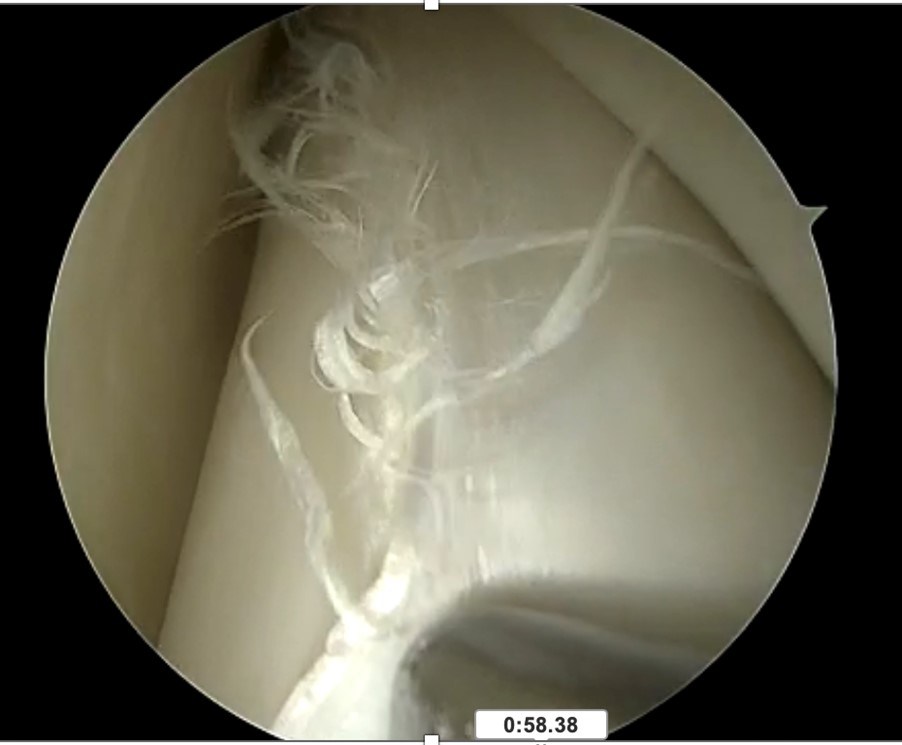
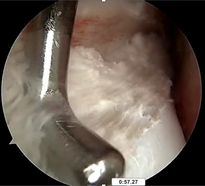
Video of the tenoscopy indicating the extent of the tear extending from under the dorsal part of the manica flexoria proximally courses laterally as a lateral marginal tear within the fetlock canal to then course dorsally again within the proximal pastern region as depicted by the sMRI and Vision CT.