

Septic pedal bone osteitis
History
This horse initially presented with a chronic foot abscess and evidence of septic pedal osteitis on radiographic examination.
Clinical Examination
After initial attempts at medical management, the horse underwent CT-guided debridement of the infected distal phalanx via the sole and a dorsal hoof wall resection. The horse continued to show marked lameness and it was decided that further information was needed to make a plan going forward.
MRI Findings
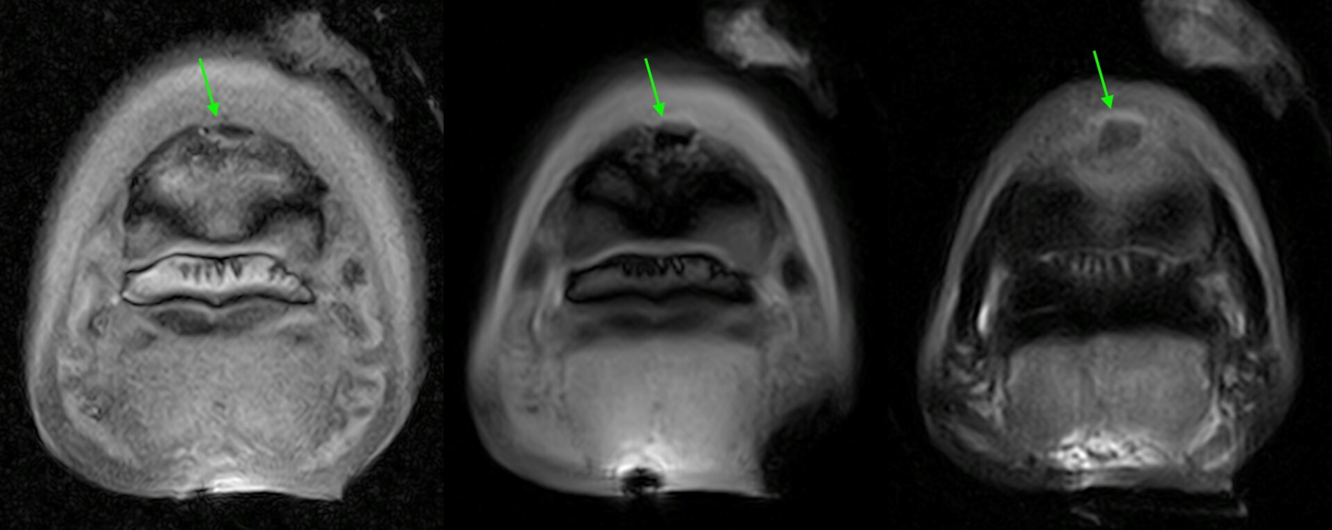
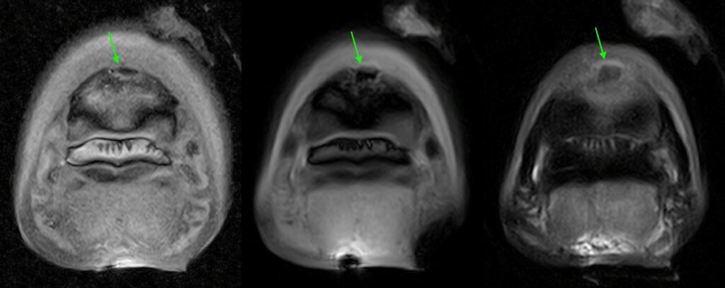
Standing Equine MRI was then performed which allowed identification of a previously unseen bone fragment likely indicating a sequestrum (See Figure 1), or piece of necrotic bone. This was confirmed at repeat surgery and removed. The horse remained in the hospital for another week for medical management but was discharged soon after.
Conclusion
MRI in this instance was vital for the management of the case. Interestingly the sequestrum was very conspicuous on the MRI, but four days prior it was not visible on the CT (even looking retrospectively). This may have been due to the rapid development of this lesion or the higher contrast resolution of MRI and the fact that the fragment is highlighted by a rim of fluid signal on MRI.
Many thanks to the RVC Equine Team for sharing this case study.
Got an MRI or CT case study you’d like to share? If so, get in touch with a summary and we’ll do the rest!


