

Canine optic neuritis
The Patient
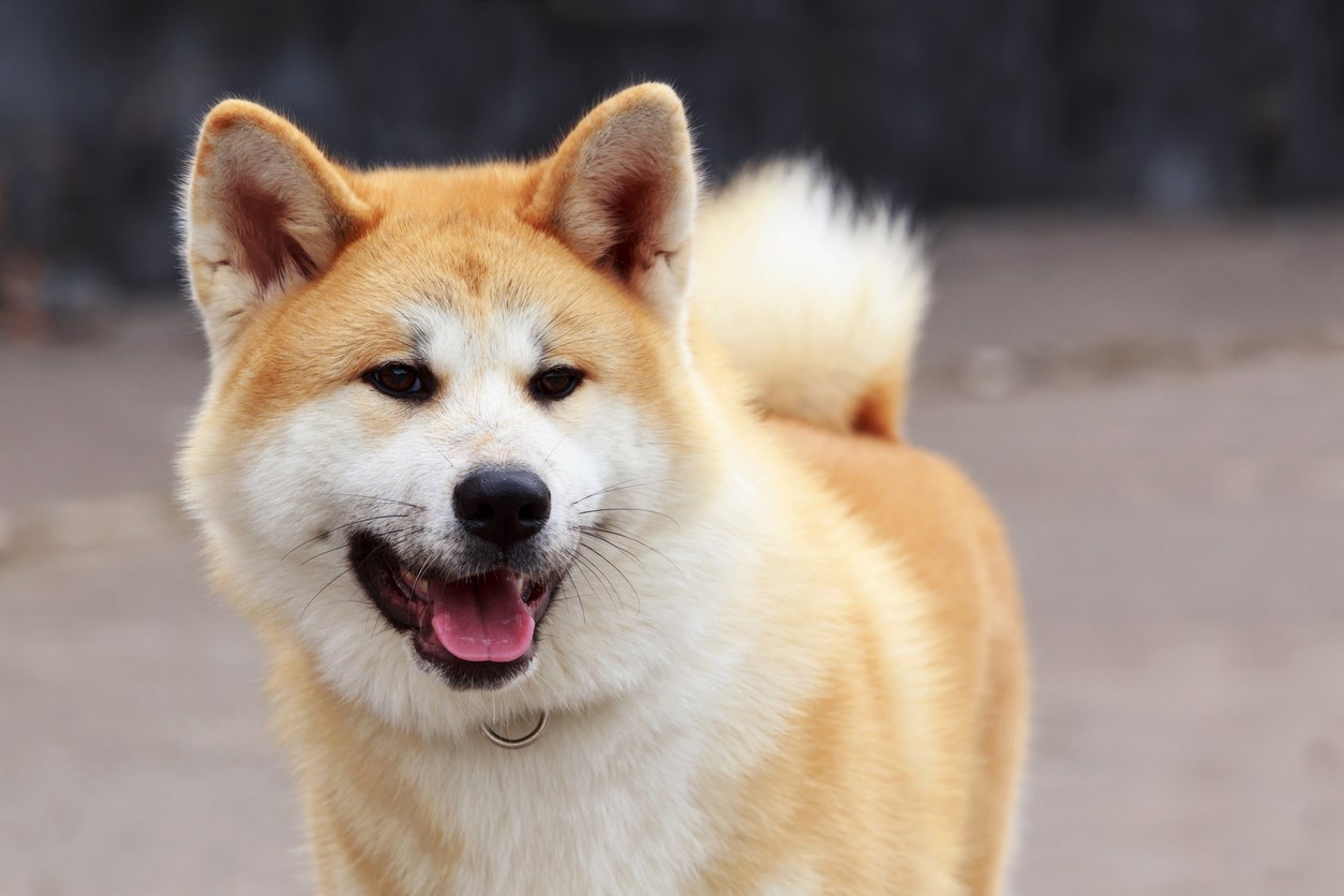
In this case study we take a look at a 5-year, 8-month-old, female Akita who presented with canine optic neuritis or acute onset blindness. Otherwise considered healthy, the dog underwent an MRI using the Hallmarq small animal 1.5T MRI machine.
The imaging results
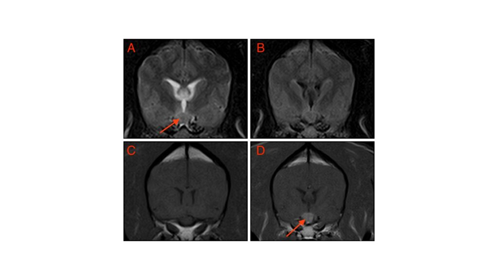
(A) T2W, (B) FLAIR, (C) T1W pre-contrast, and (D) T1W post-contrast images of the forebrain identify a relatively well-defined enlargement of the caudal optic nerves / optic chiasm. The lesion appears homogenously T2W and T1W isointense with notable homogenous enhancement (A and D arrow). There is a mild mass effect associated with this lesion which causes minimal overlying parenchymal compression. See Figure 1 below:
The most valuable MRI sequence for this case
The T1-weighted post-contrast sequence highlights the enlargement of the bilobed chiasm. Although a neoplastic process cannot be ruled out from these images, the study supported a diffuse bilateral optic nerve disease most compatible with inflammation or optic neuritis.
Optic neuritis – a disease overview
Optic neuritis, although relatively uncommon, is an important aetiologic diagnosis of acute blindness in dogs as this disorder can often be effectively treated and vision restored. Optic neuritis in dogs is usually a manifestation of meningitis of unknown origin (MUO) such as granulomatous meningoencephalitis (GME). This is an immune-mediated disorder of the central nervous system (CNS) that most commonly affects small to mid-sized young adult dogs.
The clinical manifestations are usually limited to acute vision loss and afferent pupillary defects but if the disease is more multifocal, other clinical manifestations of intracranial disease may be seen. The diagnosis should be achieved quickly to ensure minimal permanent optic nerve damage. The essential diagnostic evaluations include a complete ophthalmoscopic examination, an electroretinogram, and cross-sectional imaging with the MRI.
Optic neuritis – the prognosis
Vision can return within days of instituting immuno-suppressive therapy. However, in a recent study of 72 cases with follow-up, 50 remained blind, 10 had partial visual improvement, and 12 were assessed as having normal vision in the affected eye(s). If vision returns, it can often be maintained but sometimes continued immunosuppressive is necessary for life.
References
- Grahn BH, Starrack G, Bauer B. Diagnostic ophthalmology. Can Vet J. 2012 Nov;53(11):1223-4
- Muñiz Moris L, Cherubini GB, Caine A. Low-Field Magnetic Resonance Imaging Findings in 18 Dogs With Presumed Optic Neuritis. Front Vet Sci. 2021 Jan 7;7:585828
- Smith SM, Westermeyer HD, Mariani CL, Gilger BC, Davidson MG. Optic neuritis in dogs: 96 cases (1983-2016). Vet Ophthalmol. 2018 Sep;21(5):442-451


