

History
A horse presented with right forelimb lameness, which improved following a palmar digital nerve block. Radiographs revealed a small lytic lesion at the proximomedial aspect of the middle phalanx, although the clinical relevance of this finding was uncertain. The horse also showed a positive response to intra-articular anaesthesia of the distal interphalangeal (coffin) joint. The joint was treated with corticosteroids, but the lameness persisted despite two weeks of rest.
MRI findings
Alongside other findings, standing MRI revealed an osseous cyst-like lesion in the dorsal aspect of the medial glenoid cavity of the middle phalanx (Figs 1, 2A and 2B), with a small communication towards the articular surface of the proximal interphalangeal joint. There was mild surrounding STIR hyperintensity, consistent with bone oedema-like signal.
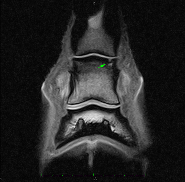
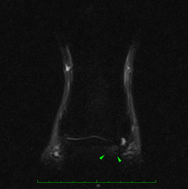
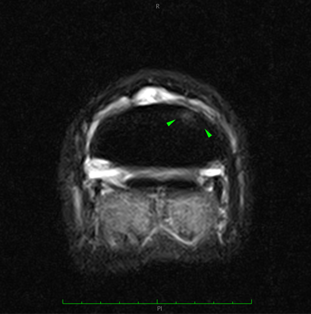
Both images show bone oedema-like signal in the medial glenoid of the middle phalanx.
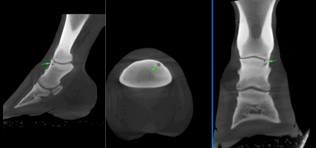
CT findings
A CT scan using Hallmarq’s Vision CT provided a clear visualisation of the cyst-like lesion and the associated indentation in the articular surface.
Conclusion
The combination of standing MRI and Vision CT provided a thorough assessment of the pathology. MRI revealed the bone oedema-like signal, while CT offered detailed structural information and clear characterisation of the cystic lesion. This case highlights how combining both modalities allows for a more comprehensive evaluation and supports a more accurate prognosis.
With thanks to Christine Svanholm, Hästklinik Florian L AB, Sweden, for sharing this case with us.


