


Computed Tomography (CT) is a powerful imaging modality adapted from human healthcare for use in veterinary medicine, including equine medicine. In this comprehensive guide to veterinary CT, we take a look at how CT can be invaluable to equine practice and the diagnosis of complex conditions.
CT provides detailed 3D imaging of structures such as the head, neck, and limbs, providing clear visualization by eliminating superimposition of tissues. This capability makes CT invaluable for investigating complex conditions, surpassing the diagnostic limitations of conventional imaging methods like radiography and ultrasonography.
Traditional radiography produces two-dimensional representations of three-dimensional structures, leading to overlapping of soft tissue and bone, which can obscure important details in anatomically complex regions.
While ultrasonography excels in soft tissue evaluation, it struggles to visualize structures deep to bone or gas-filled areas and is limited to assessing the surfaces of bones.
CT overcomes these limitations by utilizing X-ray radiation to capture multiple projections around the axis of the region of interest as the scanner rotates. These projections are processed to create a high-resolution, 3D grayscale image of the anatomy. Digital manipulation of these images enhances tissue contrast, enabling precise visualization of both soft tissue and bone.
This advanced imaging technology supports veterinarians in formulating targeted and effective treatment plans, ultimately improving outcomes for equine patients. By providing clarity and diagnostic accuracy, CT has become an indispensable tool in equine veterinary care, advancing the standard of diagnostics and treatment planning.
What is a CT Scan for Horses?
CT is an advanced diagnostic imaging technique that uses X-ray radiation to generate detailed 3D images of a horse’s anatomy. As the scanner rotates around the region of interest, it captures multiple thin slices, which are reconstructed into a three-dimensional image. This process takes only a few minutes, offering rapid and precise visualization of complex anatomical structures.
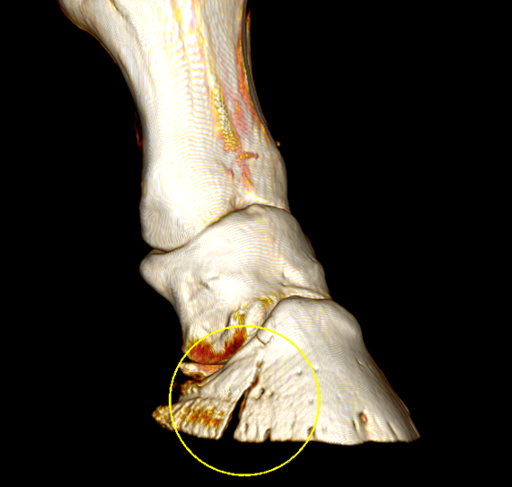
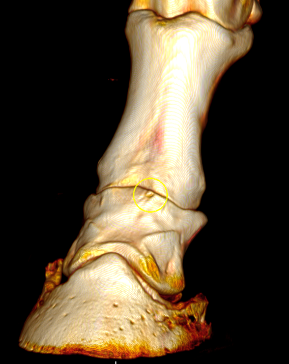
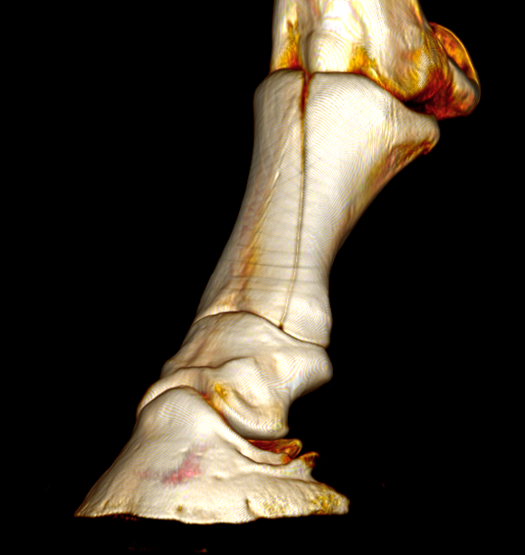
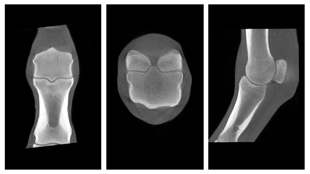
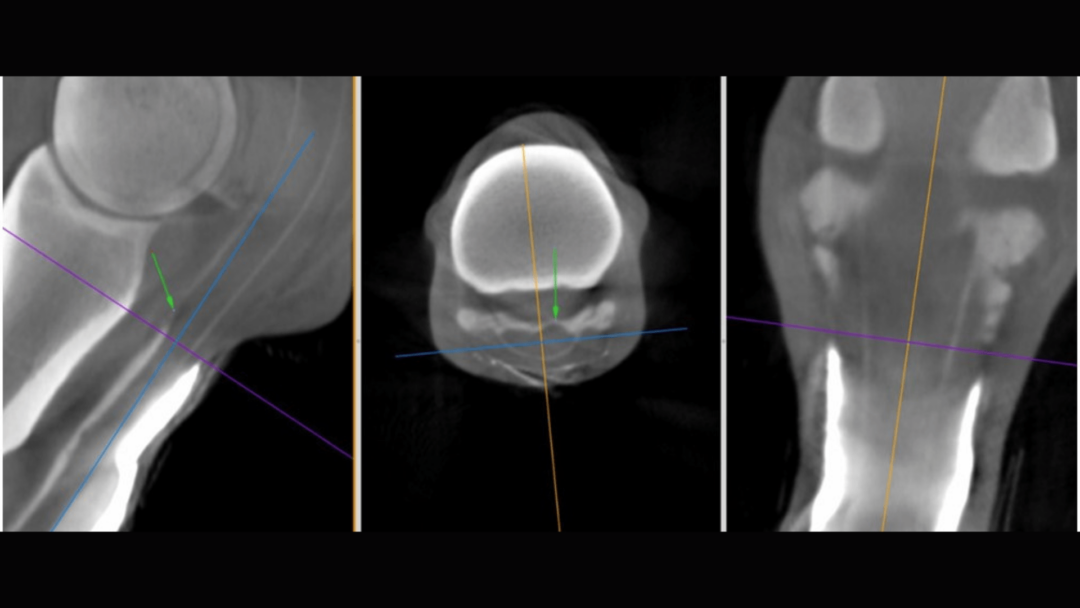
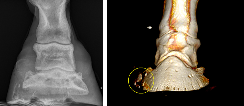
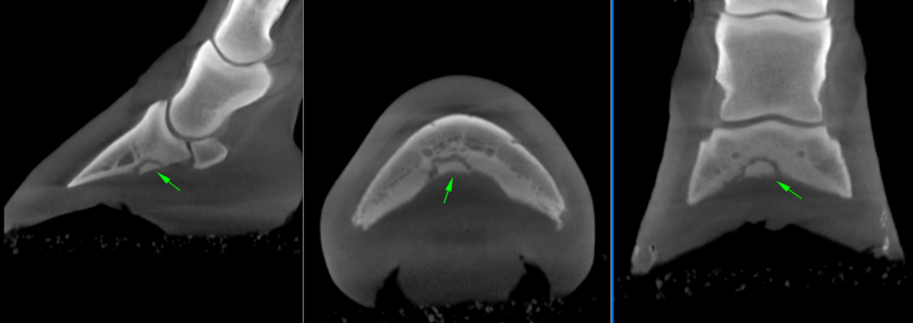
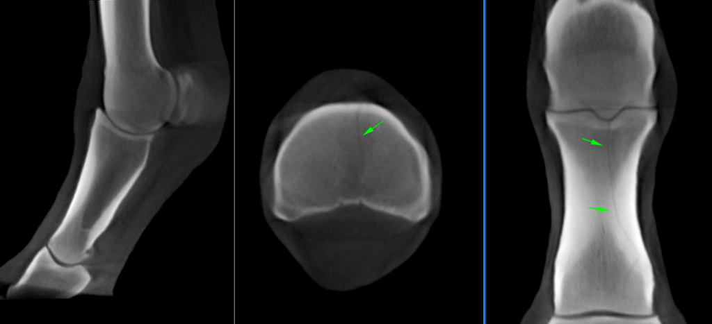
All 3D volume renders (above) taken with the Hallmarq Vision CT system.
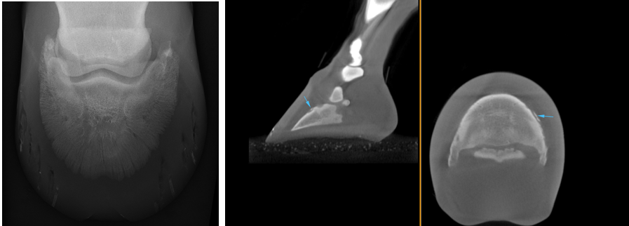
In equine musculoskeletal imaging, CT excels at assessing bony abnormalities, such as fractures and subtle changes in bone density, and is widely regarded as the gold standard for bony pathologies.
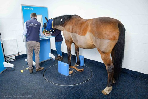
Veterinary CT systems are designed to accommodate either standing sedated horses or those under general anesthesia, with standing configurations reducing risks and costs associated with anesthesia. The short acquisition times further minimize motion artifacts, enhancing diagnostic reliability and efficiency.
Veterinary CT systems are categorized based on the geometry of their X-ray beam into fan-beam and cone-beam designs. Both systems provide comparable evaluation of bone; however, fan-beam CT offers superior soft tissue resolution.
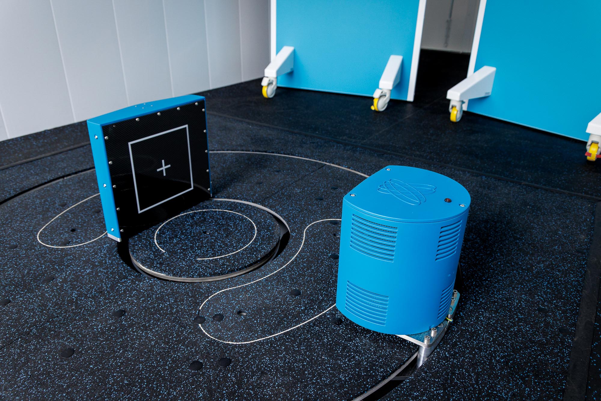
Cone-beam CT, such as Hallmarq’s Vision CT, utilizes a cone-shaped X-ray beam to efficiently capture volumetric data in a single rotation. This design enables rapid, high-resolution imaging and is particularly suited for standing horses, offering a non-invasive, practical, and cost-effective solution for accurate diagnosis and treatment planning in equine veterinary care.
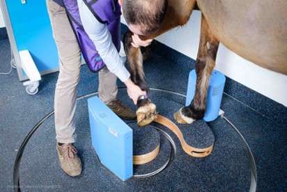
Preparing Your Horse for a Standing CT Scan
Proper preparation is key to ensuring the best results from a standing CT scan. While imaging the distal limb typically takes just 2–5 minutes—significantly quicker than an MRI—the time spent preparing the horse is invaluable.
Your veterinarian will collaborate with the referral practice prior to the scan, sharing essential case details. This includes evaluating the lameness pattern on the day of the scan, which helps contextualize the findings. It is also crucial for the veterinarian to assess and evaluate the horse’s feet before the scan.
- The horse’s shoes should be removed, as metal artifacts from the shoes can interfere with the CT motion correction software.
- Most practices take one or two X-rays before the CT scan to check for large, clenched fragments, which could also affect image quality.
- The horse’s leg should be clean and dry.
- Upon arrival at the hospital, the horse should be given time to settle in the stall, urinate, and relax before entering the scan room.
- Once in the scanning room, the goal is to keep the horse standing still while the scanner rotates around the limb to capture the images. Each scan takes around 90 seconds. Depending on the horse’s movement, a few additional scans may be necessary to achieve optimal results.
Procedure Overview and Safety Considerations
As an extremely safe diagnostic imaging modality, standing leg CT poses no risk to the patient. However, good protocol both before and during the scan can help keep the procedure to a minimum without any stress to the horse:
- Your horse will not require general anesthesia but a small amount of gentle sedation will be administered to help it remain calm throughout the procedure.
- An experienced veterinarian will choose the appropriate type of sedation based on the individual patient’s health condition and the duration of the scan.
- They will closely monitor the horse’s vital signs throughout the procedure to ensure safety.
- Once prepared for scanning, the sedated horse is walked into the CT room.
- The horse will be positioned square. This is vital to reduce motion as the horse will remain unconstrained throughout the process.
However, even the calmest horse may sway slightly while standing, causing subtle changes in joint positioning as they shift their weight. To address this, Hallmarq’s advanced motion correction software is specifically designed to compensate for these minor movements during the imaging process, ensuring precise and reliable results.
With a short acquisition time of just 90 seconds per area, the system minimizes the impact of motion artifacts. In some cases, additional scans may be required to achieve optimal results, but the entire process typically takes only 15 minutes to 1 hour, ensuring efficiency and accuracy for both horse and veterinarian.
What Equine CT Scans Can Reveal
CT has become an essential tool in equine diagnostics, enabling detailed evaluation of critical areas such as the head, cervical spine(neck), and limbs. By providing high-resolution, cross-sectional images, CT allows veterinarians to investigate complex conditions such as fractures, joint abnormalities, head and spinal pathologies with unparalleled precision. This advanced imaging modality overcomes the limitations of traditional radiography and ultrasound, which are often unable to visualize deeper or overlapping structures adequately.
With its capability to deliver rapid and accurate diagnostics, CT is instrumental in formulating effective treatment plans for orthopaedic and neurological conditions in horses.
Cranial Structures
CT is an invaluable tool for diagnosing and characterizing diseases of the equine skull, including conditions affecting the nasal passages, paranasal sinuses, and dentition. It effectively documents pathological changes in dental, osseous, and soft tissues, contributing to a growing understanding of these conditions. CT has been particularly beneficial in advancing the diagnosis of cheek tooth abnormalities, especially when accompanied by infectious or inflammatory complications such as osteomyelitis and sinusitis.
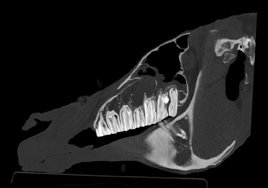
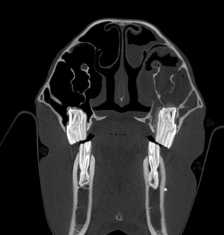
Figures 1 and 2 – 3D reconstructed CT images of a horse’s head with a tooth infection and secondary sinusitis.
Pictures courtesy of Dr. Maty Looijen.
By providing detailed imaging of the internal structure of teeth and their surrounding tissues without the superimposition issues seen in traditional radiography, CT has significantly enhanced the accuracy and depth of equine cranial diagnostics. Additionally, it offers a more comprehensive evaluation of mass lesions in the nasal passages and paranasal sinuses, such as ethmoid hematomas and neoplasia, which can be challenging to fully assess with tools like sinoscopy, endoscopy, or radiography
Cervical Spine (neck)
Veterinary CT has significantly advanced the evaluation of the equine cervical spine, particularly in cases of poor performance, neurologic deficits, or unlocalized lameness. Unlike radiography, which is limited by superimposition and muscle mass, CT provides 3D imaging that allows comprehensive assessment of the horse’s spinal column in any plane.
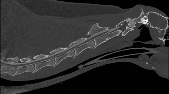
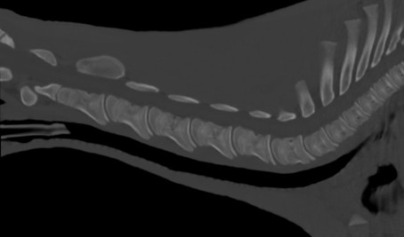
It has proven effective in diagnosing conditions such as spinal cord compression, subarachnoid space narrowing, osteoarthritis, intervertebral disc disease, and articular process abnormalities. These findings are often associated with clinical signs like neck pain and unresolvable forelimb lameness, making CT an invaluable tool for detecting and understanding complex cervical spine pathologies in horses.
Bony Pathology
Lameness is a prevalent issue in horses, often necessitating equine veterinarians to pinpoint its source and provide treatment to restore soundness. When traditional imaging methods like radiography and ultrasonography fail to identify the underlying cause, advanced diagnostic imaging, such as CT, becomes a valuable tool.
One significant advantage of veterinary CT, as with other cross-sectional imaging modalities, is the reduction of superimposition and the ability to provide more detailed visualization. CT is particularly effective in evaluating bony structures and is considered superior to magnetic resonance imaging (MRI) for assessing cortical bone. It offers greater detail of the bones in the foot compared to radiography and, in some cases, MRI.
Although MRI excels in soft tissue visualization, CT can provide useful information about soft tissues when paired with contrast agents, such as intra-arterial or intrathecal contrast, to enhance the detection of flexor tendon lesions in the distal limb. CT is also valuable for identifying foreign bodies within the hoof wall and aiding in partial hoof wall resection for conditions like keratomas.
In cases of fetlock region lameness, CT is superior to radiography in diagnosing bone and articular cartilage lesions, including sclerosis, resorption, bone marrow abnormalities, osteophytosis, fractures, and cartilage damage. Many such injuries are thought to result from cumulative damage rather than single events. Early detection through CT or MRI could help prevent severe or irreversible damage while enabling prompt and accurate diagnoses. Veterinary CT can also identify conditions such as subchondral bone disease, subchondral bone cysts, degenerative joint disease, and fractures in the tarsus and carpus
While distal limb CT is more common, CT imaging of the stifle has proven beneficial, especially with the use of contrast agents. Contrast-enhanced CT improves the detection of enthesopathy, and subchondral bone disease compared to radiography. CT arthrography, which involves injecting contrast material into the joint, is particularly useful for diagnosing intra-articular soft tissue injuries in the stifle, including meniscal damage, ligament desmopathy, cartilage injuries, and cruciate ligament pathology.
Emergency Conditions
One of the most significant advantages of veterinary CT is its rapid scan speed, which determines the time required to generate diagnostic images. This feature is particularly valuable in equine emergencies, enabling timely and accurate assessment of critical injuries. CT allows for the quantitative evaluation of fractures in the head, cervical spine, and limbs, even in the presence of casts or bandage material.
Additionally, CT is highly effective in characterizing and localizing foreign bodies, such as those from penetrating injuries or lacerations to the hoof. Within a very short timeframe, CT provides detailed quantitative assessments complemented by three-dimensional reconstructed images. These capabilities facilitate efficient decision-making, enhance surgical planning, and contribute to improved clinical outcomes in emergency settings.
The Costs of Veterinary CT
The cost of a veterinary CT scan can vary significantly depending on several factors, including:
- whether the procedure is performed under general anesthesia
- the type of CT system used
- the number of areas scanned
- the specific purpose of the imaging
As an advanced diagnostic tool typically utilized in referral centers, veterinary CT carries a higher financial cost compared to conventional imaging methods and should not be used in isolation. Effective collaboration between the primary veterinarian and the referral centers providing advanced imaging is essential to optimize its use and ensure the best outcomes for the patient.
The Benefits of Veterinary CT
Equine CT offers several significant benefits in veterinary diagnostics. By capturing high-resolution, cross-sectional images, CT provides unparalleled detail of a horse’s internal anatomy, enabling early detection of complex conditions. This capability is particularly critical in identifying fractures, bony abnormalities, and joint pathologies. Additionally, CT’s ability to generate three-dimensional reconstructions aids in surgical planning by offering precise anatomical insights, which enhances procedural outcomes and decision-making efficiency.
Beyond diagnostics, CT’s rapid imaging times, minimizes stress and risk to the patient while delivering high-quality results. Its ability to clearly visualized structures that overlap makes it indispensable for precise diagnosis and treatment. By overcoming the limitations of conventional imaging, CT contributes significantly to improved care standards and prognostic accuracy for equine patients.
Limitations of Veterinary CT
As with all imaging modalities, veterinary CT does have it’s limitations which should be taken into consideration:
- The weight and size of equine patients present challenges with the table, gantry size, and the power of the radiation generator, which prevent full body scans like those in small animals. As a result, the areas typically scanned on an adult horse are limited to the head, neck, and limbs.
- Soft tissue visualization can be poor with certain systems, especially with cone-beam CT. Fan-beam CT systems offer better soft tissue imaging, particularly with contrast agents. However, it is not as sensitive as MRI.
- Patient movement in standing CT can lead to motion artifacts, compromising image quality.
- Some CT systems introduce unique artifacts that affect image clarity.
- Radiation exposure can be a concern for personnel acquiring the images, particularly when repeat scans are needed.
Advantages of Using Veterinary CT Alongside MRI
The debate persists about which imaging modality to prioritize when the pathology is unknown. This is particularly the case in distal limb cases where multiple advanced imaging options are available. The choice often hinges on clinical presentation, suspected pathology, and logistical considerations.
While integrating CT and MRI offers a comprehensive diagnostic approach by combining the structural detail of CT with the soft tissue resolution of MRI, it is not always a financially viable option. However, with the advent of Vision CT and standing MRI systems, this dual-modality approach has become more accessible for evaluating the distal limb, providing a practical solution that balances cost and diagnostic precision.
Integrating CT with standing MRI offers a synergistic diagnostic approach by combining the distinct strengths of each modality. CT provides unparalleled visualization of bony anatomy, making it ideal for assessing cortical bone, subchondral structures, and the hoof wall. Its ability to capture high-resolution, three-dimensional images is invaluable for detecting fractures, sclerosis, and other structural abnormalities. Indeed, recent research [1] revealed that:
The combination of both CT and MRI examinations provided the most complete evaluation of bone and joint pathologies.”
In contrast, MRI excels in soft tissue evaluation, offering superior contrast resolution to identify tendon and ligament injuries, as well as bone marrow lesions (bone oedema). This dual-modality approach ensures a more comprehensive assessment by combining the physiological data from MRI with the structural detail of CT.
The Role of Hallmarq in Equine CT
Hallmarq Veterinary Imaging revolutionized equine diagnostics with the introduction of the world’s first and only Standing Equine MRI machine, a non-invasive imaging technology with diagnostic accuracy in over 90% of lameness cases. Standing Equine MRI has become a cornerstone in veterinary medicine, particularly for identifying soft tissue pathologies, which account for approximately 70% of cases seen in clinical practice by Hallmarq customers.
However, some bony pathologies can remain undetected by MRI, highlighting the need for complementary imaging modalities. Hallmarq’s Vision CT addresses this gap as a cone-beam CT system specifically designed for standing, sedated horses. This innovative technology provides high-resolution imaging of bony structures while ensuring safety, minimizing stress, and making advanced imaging more accessible. Whether used in conjunction with Standing Equine MRI, or as a standalone modality, Vision CT expands diagnostic capabilities offering a safe, effective and affordable modality for precise lameness diagnosis.
Why Choose CT Over Other Imaging Modalities?
Conventional imaging techniques, such as radiography and ultrasonography, have limitations, particularly when evaluating complex regions like the equine head, sinuses, and joints. Radiographs are two-dimensional, which can result in overlapping structures, making it difficult to visualize internal anatomy. They also fail to detect subtle bone changes until significant mineral loss occurs. Diagnostic ultrasound, while excellent for soft tissue imaging, struggles with structures deep to bone or gas-filled organs, and it is limited to evaluating only the surfaces of bones.
These limitations highlight the need for advanced modalities like CT, which provides high-resolution, cross-sectional images that allow for clearer differentiation of both bone and soft tissue, overcoming many of the constraints of conventional imaging
Comparing Veterinary CT and MRI
CT excels over MRI in diagnosing bony pathologies, offering superior resolution and detailed imaging of the bone structure, including fractures and subtle changes in bone density. It is particularly useful for evaluating complex fractures and cortical bone, providing better contrast for bony abnormalities than MRI.
MRI, on the other hand, is generally superior for soft tissue imaging, including tendon, ligament, and cartilage evaluation, but is less effective for detailed bone imaging. Both modalities have their respective strengths, and the decision between them typically depends on the suspected pathology, with CT preferred for bony issues and MRI for soft tissue-related conditions.
Conclusion
Veterinary CT scans have revolutionized equine veterinary care by providing detailed, high-resolution images of a horse’s internal structures, which is essential for diagnosing complex conditions. The ability to capture detailed 3D images allows for early detection of issues, enabling veterinarians to plan precise treatments and improve overall health outcomes for horses. Investing in advanced imaging technologies like CT scanners is crucial for equine veterinary practices, as it ensures accurate diagnostics, enhances treatment planning, and ultimately contributes to better care and welfare for horses.
References
Lin, S.-T., Bolas, N.M., Sargan, D.R., Peter, V.G. & Murray, R.C. (2025) Comparison of standing cone-beam computed tomography and low-field magnetic resonance imaging findings in the equine metacarpo- or metatarsophalangeal region of standing sedated horses. Equine Veterinary Education, 37, 125–138. Available from: https://doi.org/10.1111/eve.13967
FAQs About Standing Equine Leg CT
- What is a standing equine leg CT scan?
- Does my horse need sedation for a standing CT scan?
- How long does an standing CT scan take?
- Are standing CT scans safe for horses?
- What conditions can a standing equine CT scan detect?
- How soon will I get the results from my horse’s standing CT scan?
- How should I prepare my horse for a CT scan?
CT is similar to x-ray. It uses ionising radiation to capture multiple images from different angles, combining them all to create a 3D image.