MRI is one of the most valuable non-invasive diagnostics available in veterinary medicine. The use of MRI for evaluating musculoskeletal structures and injuries is common in equine practice and is continually expanding in small animal medicine. Jennifer Brisson, DVM, DACVR, outlines how MRI provides unparalleled evaluation of soft tissue structures over all other imaging modalities. Unique in its evaluation of osseous structures with the use of specific pulse sequences, musculoskeletal MRI (MSK MRI) generally refers to studies of the appendicular skeleton.
Common Indications of MSK MRI May Include:
Soft tissue evaluation:
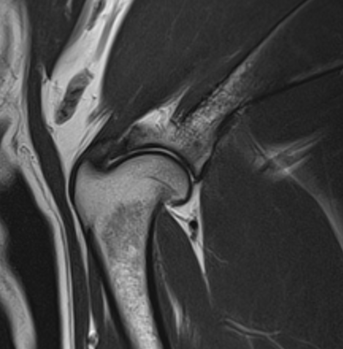
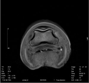
MSK MRI allows for detailed examination for injury or inflammation of soft tissue structures such as tendons, ligaments, and muscles. MSK MRI provides invaluable information when evaluating complex structures such as the canine shoulder (Fig. 1) and equine hoof (Fig. 2). Complementary to CT or radiography, tendinopathies of the supraspinatus and biceps tendons in dogs, or the deep digital flexor tendon in horses, are common diagnoses achieved with MSK MRI.

Orthopedic Conditions:
MSK MRI can provide evaluation of the articular cartilage, intra-articular structures (such as menisci), and the bones for both congenital and acquired pathologies. Indications for an MSK MRI may include acute and chronic lameness in both small animal and equine patients.
Specifically for canines, MSK MRI may be indicated to facilitate the diagnosis of developmental bone disease, such as osteochondrosis dessicans, and the extent of change within the subchondral bone. In addition, it is useful in assessing the canine stifle joint where physical examination findings are not diagnostic for cruciate ligament injury in the presence of lameness.
For equine patients, MSK MRI may be used in the diagnosis of degenerative arthopathies of the distal interphalangeal joint or lesions of the proximal phalanx.
Assessment of Neoplasia:
MSK MRI allows for lesion identification, margin evaluation, surgical planning, state of regional and sentinel lymph nodes, and response to treatment.
Treatment Monitoring:
In addition to neoplasia, monitoring of orthopedic conditions can be performed to assess response to treatment and rehabilitation and to guide interventions.
Is High-Field or Low-Field MRI Better for MSK MRI Exams?
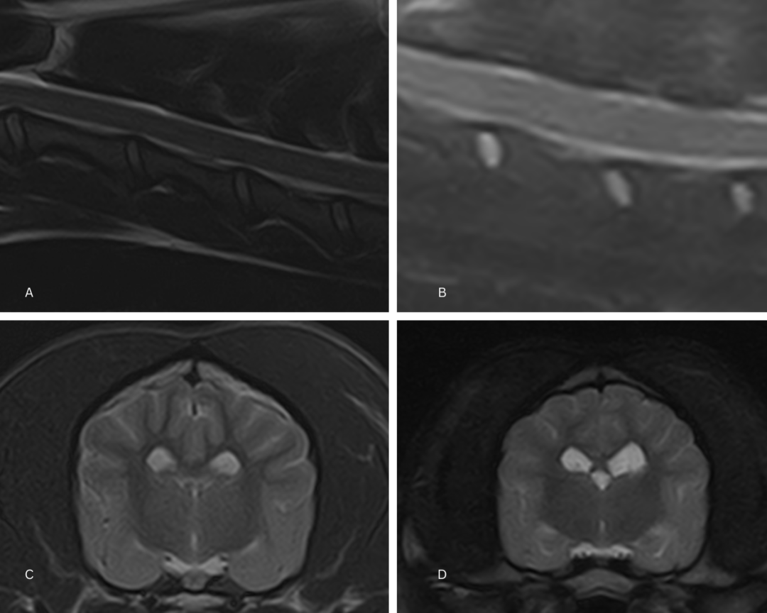
As discussed in our blog on the difference between low-field and high-field MRI, there are trade-offs to imaging with different strength magnets and indications for both. In MSK MRI, musculoskeletal tissue contrast behaves differently at different field strengths.
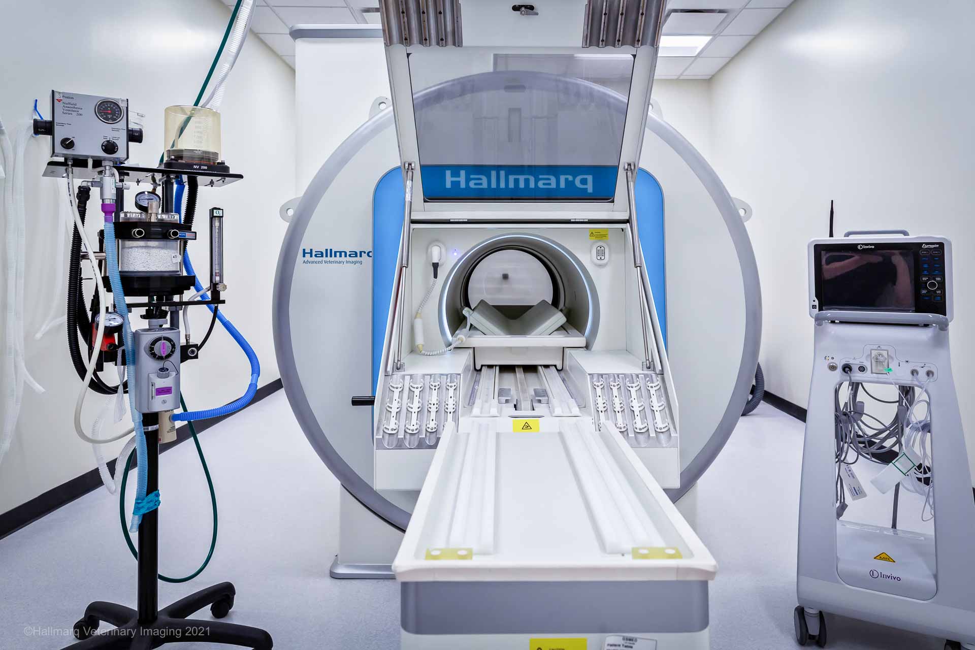
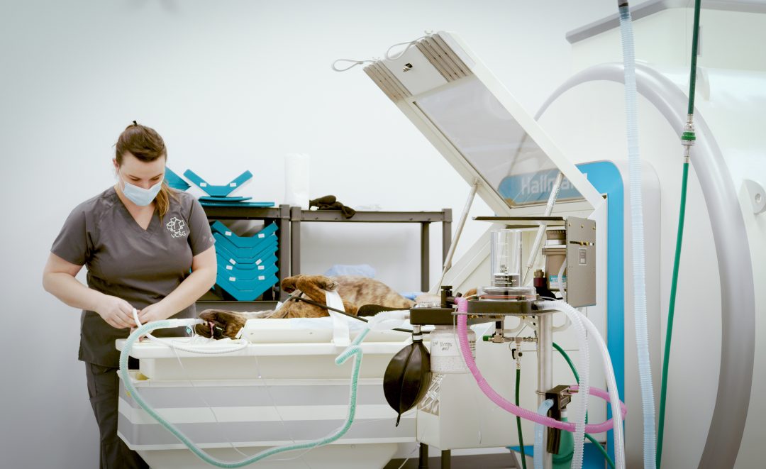
High-field MRI comes with greater magnetic field strength and generally superior image resolution. However, these exams require general anesthesia and may also come with greater expense.
Low-field MRI may be performed standing – and with sedation – and has the advantage of susceptibility artifact reduction.
The two types of MRI can even be complementary to each other in imaging the same patient.
So Why MSK MRI?
As MRI expands in availability among veterinary hospitals and specialty centers, the applications for use continue to grow as well. In cases where traditional diagnostic methods may not provide a cause for lameness, MSK MRI can offer a more comprehensive view often helping to achieve a diagnosis essential to patient care.
INTERESTED IN VISIONARY VETERINARY IMAGING?
