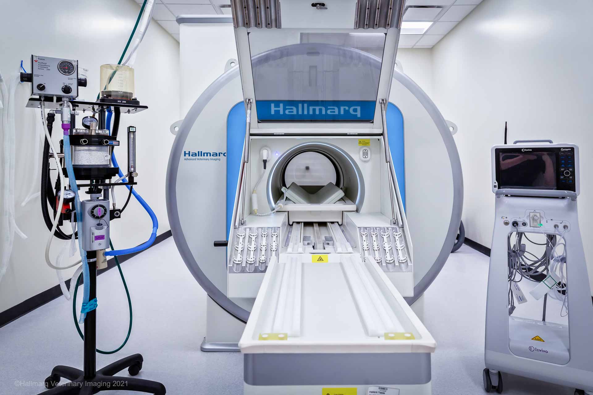
Blindness in dogs and cats can be distressing for the pet and its owner. Determining the cause of vision loss can be complex, as it can arise from various issues affecting the eye, optic nerve, or brain. Dr Simon Platt dives deeper into how Magnetic Resonance Imaging (MRI) has become a valuable diagnostic tool for veterinarians in investigating and understanding the underlying causes of blindness.
The eye, optic nerve, and brain work in conjunction to facilitate vision. Visual information is captured by the retina in the eye, transmitted through the optic nerve, and processed in the visual cortex of the brain. Any disruption in this pathway can lead to blindness. While a routine ocular examination can identify problems in the eye itself, MRI allows veterinarians to look at the eye in more detail and beyond the eye to examine the optic nerve, optic chiasm, and brain structures.
Fundic examination can be normal in cases where dogs and cats have acute vision loss secondary to ocular neoplasia which can be accurately identified on MRI. Even if a mass lesion can be visualised with fundoscopy, its physical extent requires accurate evaluation, and MRI will provide this.


The optic nerve can be involved in various conditions leading to blindness, such as inflammation, compressive lesions, trauma, and even ischaemia secondary to systemic hypertension. MRI provides detailed, high-resolution images of the optic nerve, making it easier to detect abnormalities that might not be visible through other diagnostic techniques. For instance, if the optic nerve is swollen or compressed by a tumor or fracture, MRI can identify this, aiding in diagnosis and treatment planning.
Identifying central nervous system causes
One of the key advantages of MRI when used to investigate blindness in dogs and cats is its ability to detect problems in the brain and central nervous system (CNS). When blindness is not directly caused by an issue within the eye, the CNS must be evaluated. MRI is particularly useful for identifying:
- Optic neuritis: Inflammation of the optic nerve can impair its function, leading to vision loss. Canine optic neuritis has been attributed to a focal or disseminated form of granulomatous meningoencephalitis (GME) amongst other etiologies. Magnetic resonance imaging (MRI) has been proven to help differentiate the structures within the optic nerve sheath and therefore could aid the diagnosis of optic neuritis in dogs. MRI findings in dogs with optic neuritis include contrast enhancement of the optic nerves and the optic chiasm, enlargement of the optic nerves, and changes to the optic disc. When suspected and when infectious diseases are ruled out, an immediate institution of immunosuppressive corticosteroids can help recover vision in over 50% of cases.
- Brain tumours: Masses or tumors in the brain, affecting the optic nerve, the optic chiasm, or the visual (occipital) cortex, can disrupt normal vision. MRI can provide detailed images that reveal the presence, size, and location of tumors. These can include tumours of the pituitary fossa such as pituitary macroadenomas which can be effectively treated with radiation.
- Hydrocephalus: Accumulation of cerebrospinal fluid in the brain can increase pressure on the visual pathways, resulting in blindness. MRI is effective in identifying abnormal fluid accumulation and can help with a medical or surgical treatment plan.
- Encephalitis: MRI can help detect encephalitis or meningitis, both of which can impair visual function by affecting the brain.
Conclusion
For veterinarians investigating blindness in dogs and cats, MRI is an invaluable tool. It provides a detailed look into the brain, optic nerve, and surrounding structures, allowing for the identification of underlying causes that might not be evident through standard ophthalmic exams. From detecting brain tumors and optic neuritis to uncovering inflammatory diseases and guiding treatment plans, MRI plays a critical role in improving diagnostic accuracy and treatment outcomes for pets with vision loss.
INTERESTED IN VISIONARY VETERINARY IMAGING?


