Revolutionising the approach to equine lameness examination for over two decades, MRI is still evolving to deliver detailed images that are truly transformational. AI technology can now help create magnetic resonance images that have even more clarity with enhancements to motion correction software, and with image denoising that can reduce scan times by up to 75%.
We take a look at the magic of MRI and what its continued development means for both patient and practitioner.
Why Standing Equine MRI?
Unparalleled detail across all tissue types
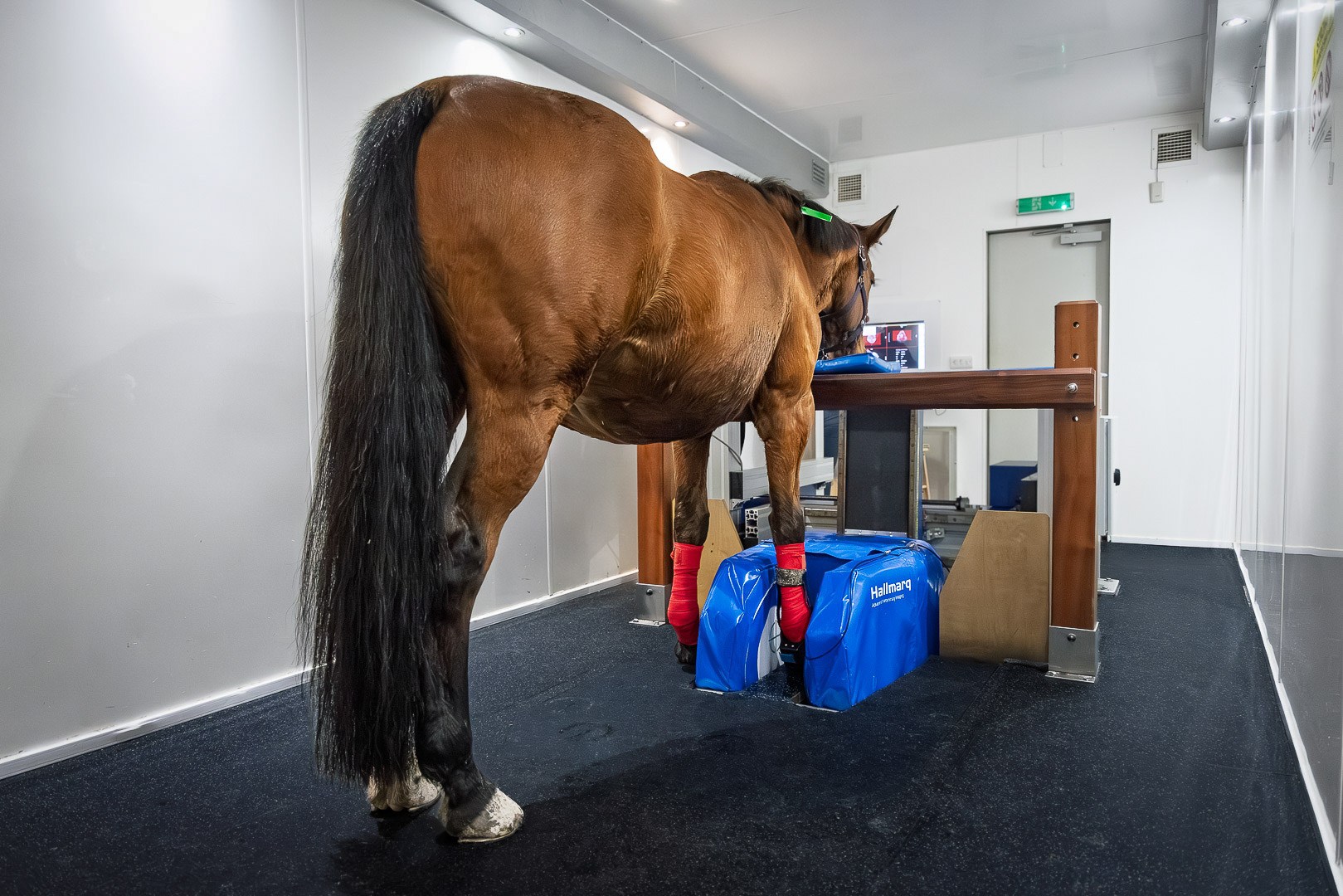
Now considered the gold standard for imaging the equine foot of the standing horse, Standing Equine MRI delivers highly detailed, three-dimensional images that visualise both bone and soft tissue.
Diagnostic in over 90% of cases, Standing Equine MRI identifies the specific cause of lameness. This level of precision is crucial for pinpointing the source of lameness, especially in the lower limb where a variety of structures can be affected. Capturing everything from the subtle tears in tendons and ligaments to the early signs of bone and cartilage changes, MRI can detect even the smallest lesion or inflammation. Its use leads to a more accurate diagnosis.
The power of prevention
MRI is not just about seeing injuries that have already happened. Its real magic is in its ability to detect early, subclinical changes: conditions like bone oedema, tendonitis, or early-stage osteoarthritis.
Often missed by other imaging techniques until they progress or become more advanced, MRI identifies these issues at their onset, allowing early intervention. Prevention is better than cure and Standing Equine MRI gives you the ability to diagnose injuries that could become more severe in the long term, giving your patients a better chance of full recovery.
Standing MRI: the safe and affordable choice
Down MRI undoubtedly places a larger demand on practice staff with highly skilled personnel needed to administer anaesthesia, and several people are required to ensure the safe positioning of the horse in the magnet. The patient will need constant monitoring for the duration of the scan process with additional costs passed on to the horse owner.
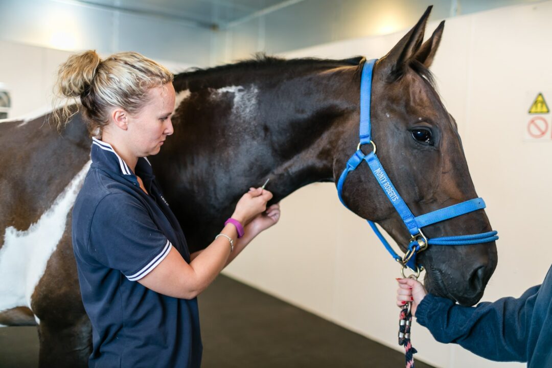
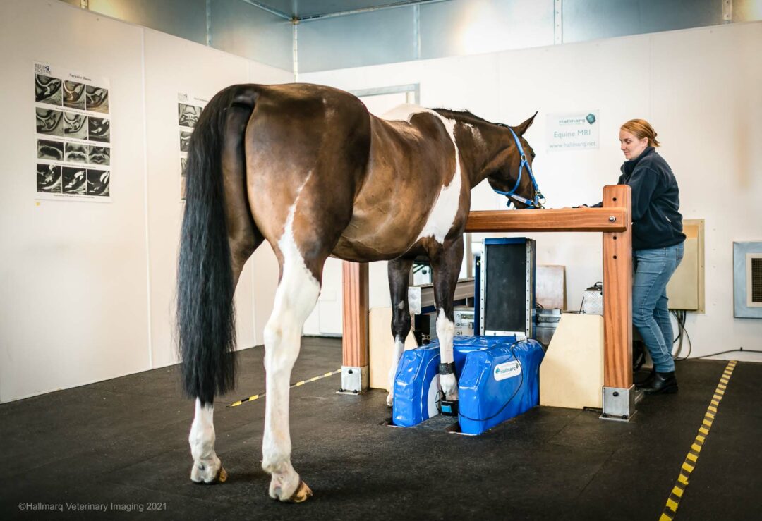
In comparison, Standing Equine MRI can be operated by just two staff – one qualified by Hallmarq to operate the machine and one to manage the patient. Since standing MRI does not require GA, mild sedation is generally sufficient, image acquisition is complete in a matter of hours as opposed to the lengthier procedure that down MRI demands. The practice and horse owner benefit from associated cost savings and most veterinarians can see more patients in less time, thus generating more revenue.

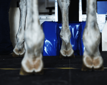
Standing Equine MRI introduces another unique difference: it allows you to image horses in their natural, weight-bearing state. This can make diagnosing acute lameness even more effective; the scan is done with the limb positioned in the way that the horse actually bears its weight. Imaging under load helps reveal problems that might otherwise only show up in a functional posture.
Precision for targeted treatment
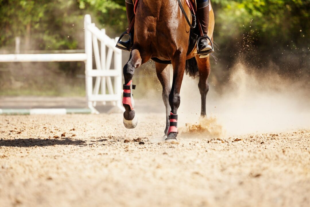
One of the biggest challenges in managing musculoskeletal injuries is determining exactly which structure within the limb is injured. With MRI, you get pinpoint precision in diagnosing the location of the injury. Whether it’s a tear in the deep digital flexor tendon, damage to the navicular bone, or early cartilage erosion in a joint, MRI gives you clear, specific answers. This level of detail allows you to tailor treatment methods with confidence, whether it’s regenerative therapy, corrective shoeing, or surgical intervention.
Seeing what other modalities can’t
In those cases where the horse remains lame – despite inconclusive findings from X-rays, ultrasound, or even nerve blocks – MRI shines. Problems like tendon or ligament injuries, bone oedema, or conditions attributed to navicular disease are often only visible with MRI.
Our comprehensive case studies gallery showcases real-life clinical cases where the use of Standing Equine MRI has made a difference in diagnosis
For example, a horse with persistent, unexplained lameness in the foot may show nothing on radiographs or ultrasound. However, MRI can reveal something like inflammation of the navicular bone or flexor tendon damage that was previously invisible.
Can we improve on the magic of Standing Equine MRI?
The answer is a resounding yes! The gold standard just got better with innovative technologies that embrace advancements in motion correction and AI algorithms to help deliver clearer images, in half the time, without any loss of resolution.
iNAV for truly transformational imaging
While scanning the weight-bearing horse has its advantages, even the most well-behaved horse will shift its weight occasionally during a Standing Equine MRI scan. Hallmarq has won industry recognition for its unique motion correction software designed to compensate for a horse’s natural sway. Now, as part of our ongoing dedication to continuously improve image quality, enhanced imaging with iNAV is available!
The latest addition to our innovative motion-correction software, this ground-breaking technology further reduces image artefacts caused by patient movement. With iNav, you can expect clearer visualisation of intricate structures, leading to more precise diagnoses.
The introduction of iNav has dramatically advanced magnetic resonance imaging of the standing horse and strengthened our confidence in diagnosing clinically significant injuries in the proximal limb.”
Dr. Natasha Werpy, DVM DACVR
iNAV is particularly beneficial for scanning anything above the foot. It can transform images of the proximal suspensory, carpus, tarsus, and fetlock regions.
Figures 1 and 2 (below) show before and after images of the proximal suspensory imaged with and without iNAV:


I have been playing with the updated iNAV. It is amazing! I was genuinely astonished by the quality of the T2W scans on a fidgety patient today. Based on the amount of motion, I was expecting the scan to look like mush, but it was beautiful. It looked better than the T1s! I’m very impressed with what you guys have done.”
Ben Anghileri, BVetMed MRCVS Clinical Director, Oakham Veterinary Hospital
iNAV has been designed specifically to further enhance what Standing Equine MRI can already see. What’s more, iNAV is available to all of our Standing Equine MRI customers and – at no extra charge! Listen to what Dr. Steve Roberts, Hallmarq’s CTO, has to say about iNAV for truly transformational imaging:
Denoising and faster scan times
Despite not needing a general anaesthetic to undergo Standing Equine MRI, it’s still better for the patient if sedation can be kept to a minimum. Less time spent in the magnet is beneficial to the horse and – from an operational point of view – less demanding on staff and practice workflow.
The AI image below shows a 75% scan time reduction. We believe AI should at least reduce scan times by 50%, with 75% possible for some of our motion correction scans. Faster scan times have the additional benefit of reduced motion.

The magic of combination scanning with MRI and CT
The operational benefits of Hallmarq’s unique Vision CT, such as avoiding general anaesthesia and minimising radiation exposure, combined with the detailed soft tissue contrast provided by Standing Equine MRI, present an impressive and comprehensive diagnostic tool. This approach reduces patient risk, enhances workflow efficiency, and provides veterinarians with a previously unattainable level of diagnostic detail.
Hallmarq customer, Dr Chris Bell from Elders Equine, explains how combination scanning – using Vision CT alongside Standing Equine MRI – gave real results that meant a faster diagnosis, the application of a targeted treatment plan, and a happy horse owner:
“Imaging with Vision CT clearly showed a complex type 2 coffin bone fracture that screening x-rays had missed, and Standing MRI enabled us to see DDFT lesions in the same patient. Combining the two modalities gave us the advantage. We’ve already seen the value of having both
Dr Chris Bell BSc, DVM, MVetSc, Board Certified Equine Surgeon (DACVS), Equine Surgery, Lameness and Sports Medicine, Elders Equine Veterinary Service, Winnipeg
Making waves with PET-MRI
Positron Emission Tomography (PET) – when used in combination with Magnetic Resonance Imaging (MRI) – has the potential to add another layer to diagnosing equine lameness making it one of the most advanced tools available in veterinary practice.
Together, PET and MRI allow you to not only see the structure of the affected limb but also understand the underlying physiological processes that might be driving the lameness.
Described by Author Mathieu Spriet DVM, MS, DACVR, DECVDI, as ‘a horse in the musculoskeletal imaging race.’ PET is seen as an actively growing field that will have a major role in the future of health care, both in human and veterinary medicine. As detailed in his recently published paper [2], this dual approach is proving invaluable for early diagnosis and targeted treatment.
What does the future hold for Standing Equine MRI?
As we look to the future of MRI, enhancing the gold standard is proving possible:
- iNAV – has transformed our cutting-edge motion-correction software, further reducing image artefacts caused by patient movement. Clearer visualisation of intricate structures leads to a more precise diagnosis.
- Denoising software – means a reduction in total scan time of at least 50% and without any loss of image resolution.
- Complementary scanning – with Vision CT means that both soft tissue and bony pathology are visible. Safe, effective, and affordable, CT offers the next logical step forward as the default for pre-surgical planning.
- PET-MRI – when combined, MRI and PET look to be taking advanced imaging to another level with the fine detail that MRI provides alongside the functional imaging of PET.
In conclusion
Unparalleled in its ability to capture every nuance, from soft tissues to hard structures, Standing Equine MRI is indispensable in the diagnosis of equine lameness examination. Its strength lies not only in what it can see but in when it can see it – often well before other modalities can. Whether dealing with subtle, early-stage conditions or diagnosing complex, multifactorial lameness, MRI is your partner in providing better, more comprehensive evaluation and care.
For veterinary teams and veterinary practice owners, MRI isn’t just another tool; it’s the magic that fills the gaps in diagnostics, supporting treatment methods tailored to each unique case.
Speak to us about the Magic of MRI

Before joining Hallmarq, Carina worked as a Technical Vet Advisor with Jurox gaining valuable commercial experience and managing two of their vacant sales territories. An equine vet for 5 years prior to this, Carina completed an internship at a busy North Yorkshire referral hospital, joining the vet team at the same practice, as well as at an equine hospital in Cambridgeshire.

Maxime has spent much of his career in agriculture-related businesses. Prior to Hallmarq, he worked IMV Technologies, l’Aigle, France where he was successively in charge of the EMEA region and then Global Key Accounts. Curious in nature, Maxime is passionate about understanding his customer’s goals and helping to achieve them.
References:
[1] Morgan, Jessica M., Helen Aceto, Timothy Manzi, and Elizabeth J. Davidson. ‘Incidence and Risk Factors for Complications Associated with Equine General Anaesthesia for Elective Magnetic Resonance Imaging.’ Equine Veterinary Journal, 7 November 2023, evj.14026. https://doi.org/10.1111/evj.14026.
[2] Spriet, M. (2022). Positron emission tomography: a horse in the musculoskeletal imaging race. American Journal of Veterinary Research 83, 7, ajvr.22.03.0051, available from: < https://doi.org/10.2460/ajvr.22.03.0051> [Accessed 21 October 2024]

