


If your veterinarian recommends spinal magnetic resonance imaging (MRI) for your pet, it’s natural to wonder how the process works and what to expect. To find out what a spine MRI is and why your pet might need one, read our previous blog article. Below is a simple, step-by-step overview of the spinal MRI procedure, from preparation to receiving results, along with an estimated timeline for each stage.
Step-by-step guide to the spinal MRI process for pets
1. Preparation (15-30 minutes)
Before the MRI, your pet will undergo a basic health check, including blood tests, to ensure it is healthy for anaesthesia; this may be done in advance of the MRI appointment. You may be asked to fast your pet for several hours before the procedure. Your vet will discuss the procedure with you and answer any questions or concerns.
2. Anaesthesia (10-20 minutes)
Since MRI requires the animal to stay entirely still for diagnostic images, the procedure is typically done under anaesthesia. This ensures your pet remains calm, safe, and comfortable throughout the scan. A veterinary team closely monitors the anaesthesia to ensure your pet’s well-being.
3. The MRI scan (30-90 minutes)
Once your pet is under anaesthesia, it is carefully placed into the MRI machine, which uses a strong magnetic field and radio waves to create detailed magnetic resonance images of the spinal cord and surrounding tissues. The duration of the MRI scan varies based on the area being imaged, the size of your pet and the complexity of the issue, but it typically takes between 30 to 90 minutes. During this time, the machine will take a series of digital images from different angles, allowing the veterinarian to assess the cervical spine in detail immediately.
4.Recovery from Anaesthesia (30-60 minutes)
After the MRI scan, your pet will be moved to a recovery area where they can safely wake up from the anaesthesia. The veterinary team will monitor your pet closely to ensure they recover well, and you’ll be informed when they’re ready to go home. Pets usually recover from anaesthesia within 30 to 60 minutes, although some may feel groggy for a few hours afterwards.
5. Reviewing the results (1-3 days)
Once the MRI is completed, the imaging report is reviewed by a veterinary radiologist, who will analyse the scans for any signs of spinal tumours, spinal cord injuries, spinal cord compression, disc issues, or other abnormalities. This process can take anywhere from a few hours to 3 days, depending on the complexity of the case and the clinic’s workload. Your vet will then discuss the findings with you and recommend the next steps based on the diagnosis.
MRI: A non-invasive and safe procedure
One of the key benefits of MRI is that it is non-invasive, meaning there are no incisions or invasive procedures involved. Your pet will not experience any pain during the MRI; the only associated discomfort issues might include mild grogginess after anaesthesia.
Safety and comfort: addressing concerns
Many pet owners worry about safety and comfort during diagnostic imaging, but it’s important to note that MRI is a safe and painless imaging technique. While the machine makes loud noises during the scan, your pet will be under anaesthesia so that they won’t be aware of any discomfort or noise. Vets and technicians are trained to ensure your pet is as comfortable and safe as possible.

MRI is a highly effective, non-invasive diagnostic tool that helps identify spinal pain and spinal issues in pets while keeping them comfortable and safe, offering peace of mind to concerned pet owners.
The only risks are related to the necessity of anaesthesia, which the patient is thoroughly assessed for so that these can be minimised.
Conditions commonly detected with spinal MRI in cats and dogs
Spinal MRI is an invaluable tool for revealing various conditions that can affect cats and dogs. It provides a high-quality image resolution of the spine, allowing veterinarians to detect and differentiate between various issues causing symptoms such as pain, mobility problems, or behavioural changes. Below are some of the most common conditions that spinal MRI can reveal:
1. Spinal tumours (benign and malignant)
Spinal tumours are one of the primary conditions that advanced imaging spinal MRI can detect in pets. These tumours can either be benign (treatable) or malignant (difficult to treat effectively), and they may develop within the spinal cord and surrounding tissues, including the vertebrae. Benign tumours, such as meningiomas, can still cause serious symptoms by compressing the spinal cord, while malignant tumours, such as osteosarcomas, can invade the spinal cord and cause more aggressive symptoms, reducing treatment options.
Spinal tumours often present with clinical signs, which can include pain, difficulty walking, incoordination of the limbs, and incontinence. MRI provides correct diagnosis with clear images that help veterinarians assess the size, location, and sometimes the type of the tumour, which is critical for determining whether surgery, radiation, or medical management are needed.
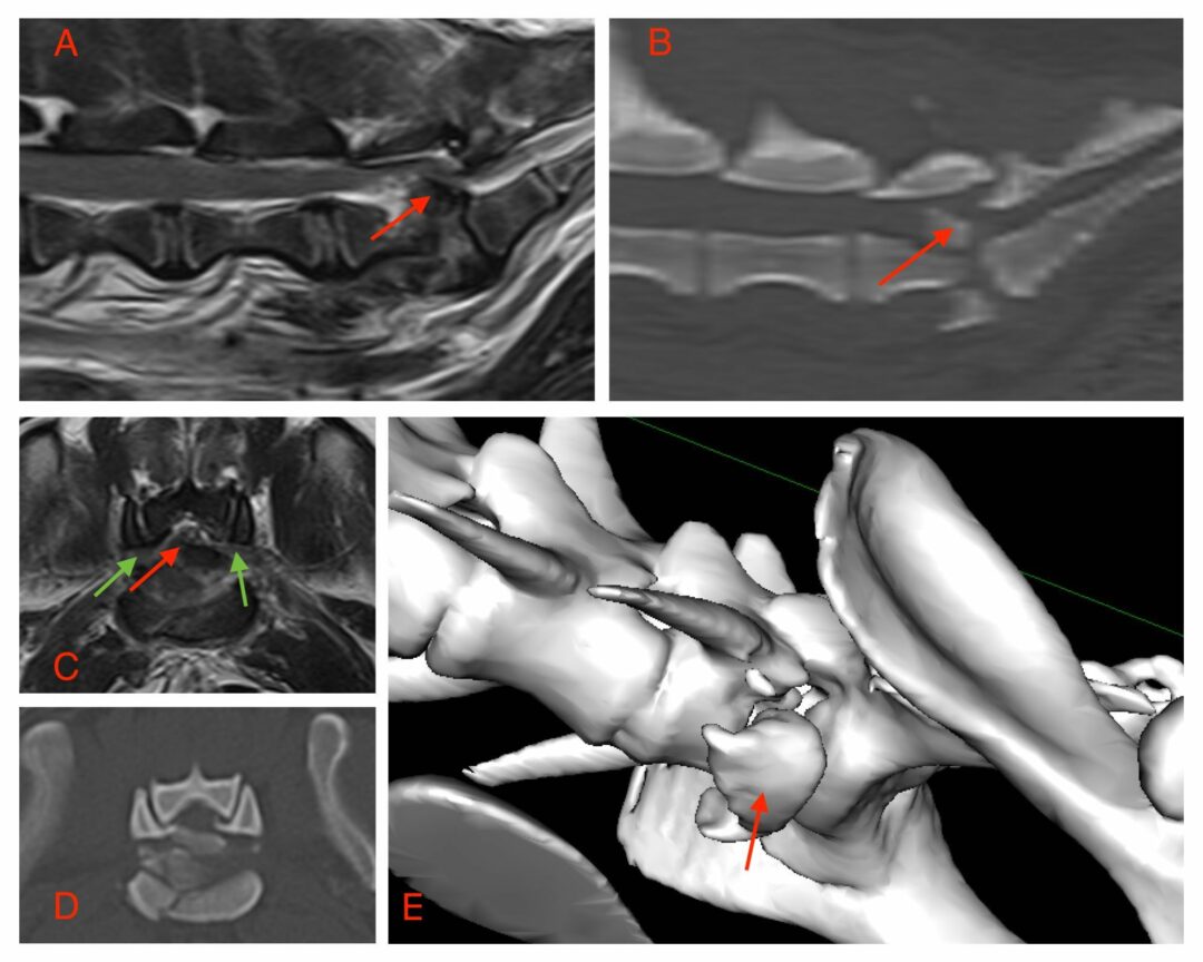
2. Herniated discs (Intervertebral Disc Disease – IVDD)
Herniated discs are another common condition in dogs; occasionally, the feline spine can mimic the symptoms of a spinal tumour. In this condition, the discs between the vertebrae become damaged or displaced, pressing on the spinal cord or nerves and causing pain, weakness, or paralysis. Certain breeds, such as Dachshunds and French bulldogs, are particularly prone to intervertebral disc disease (IVDD), where MRI is crucial in pinpointing the exact location and severity of the herniation.
MRI provides superior soft tissue imaging of the soft tissues in and around the spinal cord, making it possible to identify whether a herniated disc is causing the pet’s symptoms. This clarity ensures that the correct treatment, whether medication, physical therapy, conservative treatment, or spinal surgery, is given.

3. Spinal infections
Spinal infections, such as discospondylitis (a disease of the vertebrae and intervertebral discs), can also be detected using MRI. These infections can cause pain, fever, and mobility issues that may be mistaken for other spinal problems like tumours or herniated discs. Discospondylitis is often caused by bacterial or fungal infections, which can spread through the bloodstream to the spine and can usually be effectively treated with antibiotics.
MRI is particularly useful for detecting spinal infections because it can reveal inflammation, abscesses, or other abnormalities in the spinal tissues that might not be visible on X-rays, radiographic images, or Veterinary Computed Tomography (CT) scans. This allows for a more accurate diagnosis and helps vets determine whether drug treatment, such as antibiotics or surgery, is necessary to manage the infection.
4. Spinal cord inflammation (Myelitis)
Inflammation of the spinal cord, known as myelitis, can occur due to infections, immune-mediated diseases, or occasionally trauma. Symptoms of myelitis can include pain, weakness, and difficulty walking, which may resemble the signs of a spinal tumour or a herniated disc. In these cases, MRI is essential for distinguishing between different causes of spinal cord inflammation, as it provides detailed images of the soft tissues and reveals whether the inflammation is localised or spread throughout the spinal cord.
MRI: The key to accurate diagnosis
The detailed images provided by MRI are crucial for distinguishing between these various conditions. While some issues, such as spinal tumours, require surgical intervention, others, like infections or inflammation, may be treated with medications. The ability of MRI to reveal the precise cause of the symptoms is essential for determining the most effective treatment plan. Without MRI, many spinal conditions could be misdiagnosed or untreated for longer, potentially leading to worsening symptoms and decreased quality of life for pets.
In summary, spinal MRI is a critical diagnostic tool for detecting a wide range of conditions in cats and dogs, including spinal tumours, herniated discs, trauma, infections, and inflammation. Its ability to provide clear, detailed images helps veterinarians make accurate diagnoses and determine the best course of action to improve pets’ health and well-being.
Early detection of spinal tumours in pets offers significant advantages for their health and quality of life. Here’s why diagnosing tumours early is crucial:
1. Increased treatment options and better chances of recovery
Catching a spinal tumour early dramatically increases the number of treatment options available. In the early stages of tumour development, treatments such as surgery, radiation therapy, or even medications can be more effective in halting the tumour’s growth or removing it entirely. When tumours are small and localised, surgery is often less invasive, with shorter recovery times. Early diagnosis can also allow targeted therapies, like radiation or chemotherapy, to be implemented before the tumour spreads, greatly improving the pet’s chances of a full recovery or long-term management of the condition.
2. Enhanced quality of life
One of the key benefits of early tumour detection is the opportunity to reduce or eliminate pain for your pet before it becomes severe. Spinal tumours can cause a variety of symptoms, including pain, weakness, and even paralysis as they grow and compress the spinal cord or nerves. Early detection allows veterinarians to address the tumour before these symptoms worsen, which helps maintain your pet’s mobility, comfort, and overall well-being. Treating a tumour early can prevent severe complications, such as irreversible nerve damage, that could drastically reduce a pet’s quality of life.
By intervening early, pets can continue enjoying their daily activities, free from the discomfort an untreated tumour would eventually cause. This proactive approach helps preserve their ability to move, play, and interact without significant pain or mobility issues.
3. Reduced long-term veterinary costs
While the cost of advanced diagnostic tools like MRI might initially seem high, early detection of spinal tumours can reduce long-term veterinary expenses. By diagnosing and treating a tumour early, pet owners can avoid the potentially higher costs associated with protracted non-specific approaches, advanced-stage treatment, emergency surgeries, or long-term palliative care for severe complications. Addressing the issue early can prevent the need for more complex and expensive treatments down the line, saving money and sparing pets from prolonged suffering.
Proactive diagnostic care offers the benefit of a more affordable long-term care plan and gives pets a better quality of life by addressing issues before they worsen.
In conclusion, early tumour detection in pets increases the chances of successful treatment, reduces pain and complications, and can lower long-term veterinary costs. This makes early diagnosis through tools like MRI essential to ensuring the best possible outcome for pets diagnosed with spinal tumours.
Conclusion: prioritising your pet’s health
In conclusion, prioritising your pet’s health through imaging for assessment diagnostic tools like spinal MRI can significantly improve their quality of life and treatment outcomes. Here are the key takeaways:
- Early tumour detection: Spinal MRI enables veterinarians to identify tumours and other abnormalities early, leading to more effective treatment plans.
- Improved outcomes: By providing detailed, high-resolution images of soft tissues, MRI allows for precise diagnosis and targeted treatments, enhancing the chances of recovery.
- Better quality of life: Early detection and accurate diagnosis through veterinary MRI can help maintain or improve your pet’s quality of life by addressing issues before they become more severe.
- Safe and effective: MRI is a non-invasive, radiation-free procedure considered safe and well-tolerated by pets.
If you notice any concerning symptoms in your pet, such as changes in gait, unexplained pain, or neurological issues, don’t hesitate to consult your veterinarian for imaging in dogs or cats. They can assess whether a spinal MRI is appropriate for your pet’s specific situation and guide you through the diagnostic process.
Remember, early detection and accurate diagnosis are crucial for your pet’s health and well-being. By staying vigilant and working closely with your veterinarian, you can ensure the best possible care for your pet.