Spinal MRI: a highly effective diagnostic tool
One of the most reliable ways to detect spinal disease in pets as a cause of weakness is through spinal MRI (Magnetic Resonance Imaging). This advanced diagnostic tool provides highly detailed spine images, allowing veterinarians to visualise the spinal cord, nerves, surrounding spinal bones and their muscles. With this precise 3D imaging, veterinarians can better understand the extent of most diseases and develop the most appropriate treatment plan. Magnetic resonance imaging for pets is beneficial because it provides a non-invasive method to assess complex spinal structures that are not easily detected through traditional X-rays or CT scans.
Conditions typically discovered with spinal MRI
A spinal MRI can reveal a variety of conditions, such as:
- Intervertebral disc disease (IVDD)
- Infections and inflammation
- Spinal ‘strokes’,
- Spinal tumours
- Spinal cord compression
This makes MRI an invaluable tool in diagnosing a wide range of spinal issues in pets at an early stage.
The importance of early spinal tumour detection in pets
These tumours can cause a range of symptoms, from subtle changes in mobility to severe pain and paralysis, which, if left untreated, can drastically reduce a pet’s quality of life. By identifying spinal tumours early, veterinarians can intervene more effectively, potentially improving prognosis, reducing pain, and preserving mobility. Early detection allows for timely treatment that can prevent further damage to the spinal cord and surrounding structures, leading to a more successful outcome for the pet.
Hallmarq: leading provider of advanced veterinary imaging solutions
As a trusted leader in advanced veterinary imaging, Hallmarq offers innovative MRI systems specifically designed for animals and their unique anatomy. Their cutting-edge technology helps improve pet health and well-being by enabling early detection and accurate diagnosis. Hallmarq’s systems are tailored to meet animals’ unique anatomical and diagnostic needs, ensuring the best care possible.
In this blog, we will further explore how spinal MRIs and early detection can enhance the quality of life for your dog or cat.
Why spinal MRI is crucial for tumour detection
Spinal tumours in pets are abnormal growths that can develop within or around the spine, affecting the spinal cord, nerves, and surrounding tissues such as bones and muscles. These tumours can be benign or malignant, and their severity depends on their specific type, size, location, and how quickly they grow.
Spinal tumours can cause a range of symptoms including:
- Mild discomfort
- Weakness
- Severe pain
- Paralysis
- Loss of coordination
As the tumour grows, it may compress the spinal cord, leading to neurological deficits that impact an animal’s ability to move, stand, or walk. Spinal tumours can significantly reduce a pet’s mobility and overall quality of life without timely intervention, making early detection crucial. Veterinary diagnostic imaging plays a pivotal role in effectively identifying these conditions.
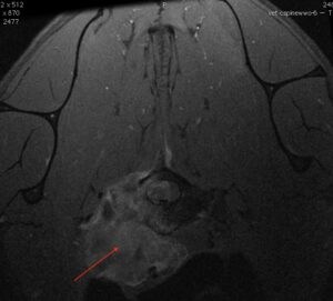
The image on the right shows a cross-sectional MRI of a vertebra clearly demonstrating a large irregular lesion associated with the vertebral body (arrow) and compressing the spinal cord. The lesion was confirmed to be a bone tumour called chondrosarcoma.
MRI vs. other diagnostic methods
MRI (Magnetic Resonance Imaging) is the most precise and effective tool when diagnosing spinal tumours and related conditions in pets. While traditional methods such as X-rays and CT (Computed Tomography) scans are valuable for identifying bone abnormalities, they often fall short in assessing soft tissues like the spinal cord, nerves, and muscles. X-rays primarily capture images of bones, making them helpful in detecting fractures or other skeletal issues. Still, they cannot provide detailed photos to assess soft tissue structures or detect small tumours.
CT scans offer more detailed images than X-rays and can show some soft tissue contrast in 3D. However, they still lack the resolution to fully visualise the intricate details of the spinal cord and surrounding tissues. In contrast, MRI provides superior soft tissue imaging, allowing veterinarians to detect even small spinal tumours and assess their impact on nearby structures more precisely. This makes MRI the gold standard for diagnosing spinal issues that involve soft tissues, including tumours, herniated discs, and inflammation.
The benefits of early detection with MRI
One of the most essential benefits of spinal MRI is its ability to detect tumours early, even before symptoms become severe. Early detection significantly improves treatment outcomes, allowing veterinarians to intervene sooner, potentially slowing the tumour’s progression and minimising damage to the spinal cord. This can help preserve an animal’s mobility, reduce pain, and improve their overall quality of life. In cases where surgery or other treatments are needed, early diagnosis through MRI gives veterinarians the information they need to plan more effective and targeted treatments.
MRI can also extend a pet’s life expectancy by catching spinal tumours in the early stages. Timely treatment can prevent further deterioration and allow pets to enjoy a longer, healthier life with fewer complications. Overall, MRI’s ability to provide detailed, accurate information about spinal tumours and surrounding structures makes it an invaluable tool for veterinarians and pet owners alike.
Signs your pet might need a spinal MRI
Spinal disease in pets can be difficult to detect early on, but specific symptoms may indicate a problem that requires further investigation. If you notice any of the following signs, it could suggest that your pet has a spinal disease, which in some cases could be a serious condition like a spinal tumour:
- Limping or lameness: If your pet is favouring one leg or seems to struggle with coordination, it may be due to nerve or spinal compression.
- Difficulty walking or stumbling: Pets with spinal problems may struggle walking, display unsteady movements, or drag their back legs.
- Sensitivity along the spine: If your pet reacts with pain or sensitivity when touched along their spine, it could signal an underlying spinal issue.
- Visible pain: Obvious signs of pain, such as yelping, whimpering, or flinching when moving, are common indicators that something is wrong with the spine or back.
Behavioural changes as early indicators
In addition to physical symptoms, behavioural changes can be early warning signs of spinal conditions:
- Reduced activity: A once playful and active pet may suddenly become lethargic or show less interest in physical activity.
- Reluctance to jump or climb stairs: If your pet hesitates to jump onto furniture, climb stairs, or engage in their usual activities, it might be due to pain or discomfort caused by a spinal problem.
- Loss of appetite or changes in mood: Some pets may experience a decreased appetite or show signs of irritability, restlessness, or depression due to spinal discomfort.
When to consult your veterinarian
If you notice any of these signs in your pet, it’s essential to consult your veterinarian. While these symptoms don’t always indicate a spinal tumour, they can point to various spinal issues that may require further investigation. Your veterinarian may recommend a spinal MRI to get a clear, detailed view of your pet’s spine and identify the root cause of their discomfort. Early detection can make all the difference in ensuring your pet receives the proper treatment and care.
Knowing when to consult your vet about a spinal MRI for your pet can be crucial in diagnosing and addressing potential spinal issues. While not every case will require an MRI, there are specific situations when discussing this diagnostic tool with your veterinarian can be essential for your pet’s health.
If your pet has been displaying signs of discomfort, such as difficulty walking, sensitivity along the spine, or changes in behaviour, it’s essential to consult your vet. During the initial evaluation, your vet will perform a physical examination and may use initial diagnostic tools, such as X-rays or blood tests, to further investigate the cause. If these tests do not provide a precise diagnosis or the symptoms suggest a problem involving the spinal cord or surrounding tissues, your vet may recommend an MRI.
How vets determine the need for MRI screening
Veterinarians typically recommend spinal MRI when they suspect a condition that cannot be fully diagnosed using other imaging methods, such as X-rays or CT scans. If your pet is experiencing symptoms like acute or chronic pain, unsteady gait, limping, or weakness that doesn’t respond to initial treatment, an MRI may be necessary to get a more detailed view of the spine. The MRI allows the vet to assess the spinal cord, nerves, and soft tissues, which are not visible on traditional X-rays. It is a powerful tool for detecting conditions like tumours, spinal ‘strokes’herniated discs, or even traumatic injuries.
When pain or discomfort persists beyond two weeks
If your pet has been showing signs of pain, discomfort, or mobility issues for over two weeks, contacting your vet’s a good idea. Ongoing pain or discomfort could indicate an underlying issue, such as a spinal problem or another neurological condition that needs further evaluation. In these cases, your vet may consider an MRI to identify the cause and develop an appropriate treatment plan.
Urgent symptoms: seek immediate veterinary attention
If your pet suddenly loses mobility, experiences severe pain, or shows dramatic behavioural changes, such as uncontrollable whining or refusing to move, you should seek veterinary attention immediately. These symptoms could be signs of a severe spinal issue, like a ruptured disc or spinal cord injury, which may require urgent intervention. In such cases, an MRI can be crucial for quickly identifying the problem, guiding emergency treatment, and providing information on the possible success of medical or surgical interventions.
By staying aware of these signs and working closely with your veterinarian, you can ensure your pet receives the appropriate care, including spinal MRI if needed, to maintain their health and well-being.
Conclusion
Spinal MRI has revolutionised veterinary medicine by offering unparalleled diagnostic imaging capabilities for various conditions. Utilising advanced tools like MRI scans and other diagnostic imaging technologies, veterinarians can detect issues in dogs, cats and other small animals, including spinal tumours and herniated discs, with exceptional precision. This advanced imaging technique uses powerful magnets to create cross-sectional and clinical images of the spinal cord tissue, providing a detailed picture of the problem.
While there may be potential risks, the benefits of early detection for conditions in dogs, including canine cancer patients, far outweigh the common concerns. With strong collaboration between the veterinary team and referral practices, diagnostic imaging tools continue to improve veterinary care, addressing various health issues and improving pet outcomes. When considering imaging options for your pet, always consult with your veterinarian to determine the best diagnostic testing approach for their unique pet needs and concerns.
In our next blog we will discuss a step-by-step guide to a spinal MRI for cats and dogs.