

Injury of the proximal metacarpal region
The patient
An 11-year-old Arab gelding endurance horse was presented with marked left forelimb lameness and mild swelling associated with the proximal palmar metacarpal region. In this case study, we review how magnetic resonance imaging (MRI) provided valuable information in aiding the diagnosis of a proximal metacarpal injury, enabling a comprehensive evaluation of both soft tissue and bone structures.
Initial examination
Ultrasonography revealed a marked increase in the size of the medial lobe of the suspensory ligament distal to its origin and loss of its normal architecture. The findings were consistent with acute desmopathy of the suspensory ligament. See Figure 1.

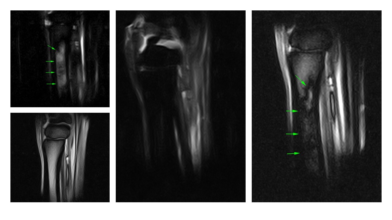
MRI findings
The horse underwent a Standing equine MRI to better evaluate the injury and fully appraise the third metacarpal bone. This revealed the presence of a marked fluid signal (“bone oedema”) originating in the region of the palmar cortex of the third metacarpal bone, distal to the proximal margin of the bone, extending distally within the diaphyseal medulla (green arrows); this was more pronounced medially. A fracture line was not clearly identified, but the degree of fluid signal was highly suggestive of a prodromal fracture of the third metacarpal bone. There was marked enlargement of the medial lobe of the suspensory ligament extending from its origin to the mid/distal third of the metacarpal region and nearly complete obliteration of the normal appearance (orange arrows).


Figures 2 and 3: T2*W and STIR sagittal MR scans of the left proximal metacarpal region (dorsal is on the left) showing the marked presence of fluid signal in the proximal metacarpal region and the metacarpal medulla. In Figure 2 there is a large region of fluid-fat cancellation artefact.


Figures 4 and 5: T1W transverse MR scans of the left proximal metacarpal region (medial is to the left). Fig 4 is more proximal to Fig 5. The orange arrows highlight the enlargement of the medial aspect of the proximal suspensory ligament.
Outcome
This horse sustained an acute injury of the suspensory ligament and third metacarpal bone. Current literature [1] suggests that injuries at the proximal metacarpal region are among the most common causes of lameness in endurance horses. The bone bruise associated with the third metacarpal bone was marked. Although the risk of propagation of a fracture was low, it could not be completely ruled out without further advanced imaging such as CT.


Figures 6 and 7: Repeat T2*W and STIR sagittal MR scans of the left proximal metacarpal region (dorsal is on the left) acquired six months after the original injury showing complete resolution of the fluid signal.
The horse had intralesional medication of the suspensory ligament with platelet-rich plasma and a follow-up standing equine MRI after six months showed complete resolution of the “bone oedema” of the third metacarpal bone. There was new thickening and sclerosis of the medial aspect of the palmar cortex of the third metacarpal bone. There was a mild improvement to the previous enlargement of the medial lobe of the suspensory ligament.
The recommendation was for the rehabilitation programme to continue, and the suspensory ligament injury to be monitored with ultrasonography.
With thanks to Dr. Giorgio Ricardi, DrMedVe, CertISELP, MRCVS, and Donnington Grove Equine Vets, UK for providing this case study.
[1] Likon, I.; Dyson, S.; Nagy, A. Magnetic Resonance Imaging Measurements of the Proximal Palmar Cortex of the Third Metacarpal Bone and the Suspensory Ligament in Non-Lame Endurance Horses before and after Six Months of Training. Animals 2023, 13, 1106. https://doi.org/10.3390/ani13061106


