In the rapidly evolving world of veterinary medicine, the integration and application of advanced diagnostic technologies have become pivotal in enhancing the care and treatment of equine patients. Among these latest advancements, combination scanning with Vision CT and standing MRI stands out as a game-changing approach to equine diagnostics. This innovative method is now being embraced by numerous centers worldwide heralding a significant leap forward in veterinary diagnostic capabilities for equine healthcare.
One of the most recent and notable adopters of this advanced diagnostic combination is The Royal (Dick) School of Veterinary Studies, The University of Edinburgh (Dick Vet). Nineteen years after its pioneering adoption of standing MRI technology, Dick Vet Equine has established itself in Scotland by offering both standing CT and MRI as a comprehensive package. Dick Vet is just one of the many centers whose dual-modality approach has ushered in a new era for distal limb imaging in horses, offering unparalleled detail and precision.
The Power of Combination Scanning
Integrating standing CT and MRI technologies provides a holistic view of equine distal limb conditions, drastically enhancing diagnostic and treatment expertise at veterinary hospitals and clinics. In particular, this rigorous combination is useful for addressing lameness and foot problems in horses, two of the most common and challenging issues faced in equine healthcare.
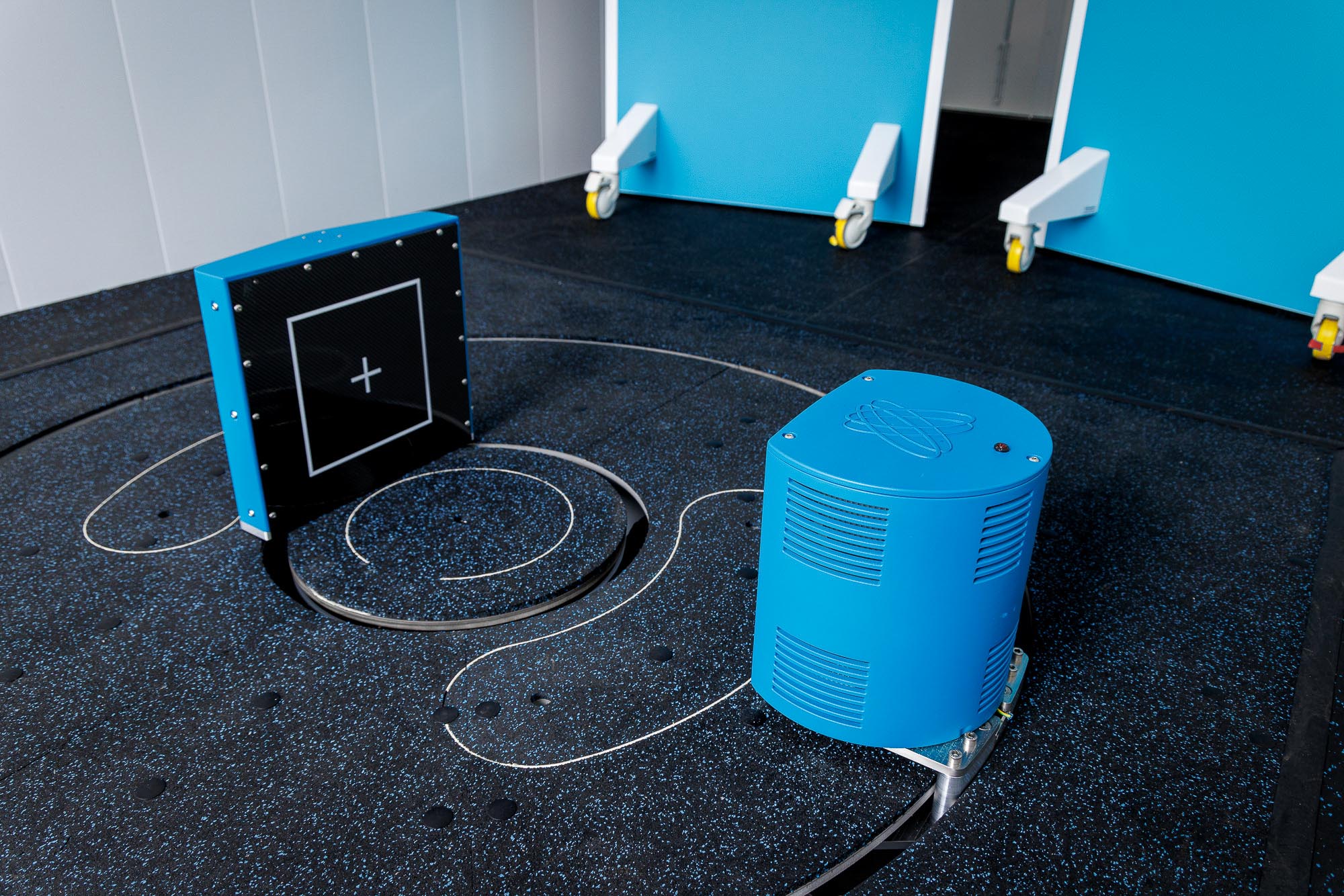
Hallmarq’s Vision CT scanner, with its state-of-the-art Cone Beam (CBCT) technology, plays a crucial role in this diagnostic revolution. Designed for precision and safety, the scanner encapsulates the leg and performs high-resolution 3D scans, taking 900 images in just a few minutes. Its ability to focus on specific areas of the lower leg minimizes radiation exposure, ensuring the safety of both patients and the veterinary professionals involved.
Innovative Design for Enhanced Safety and Efficiency
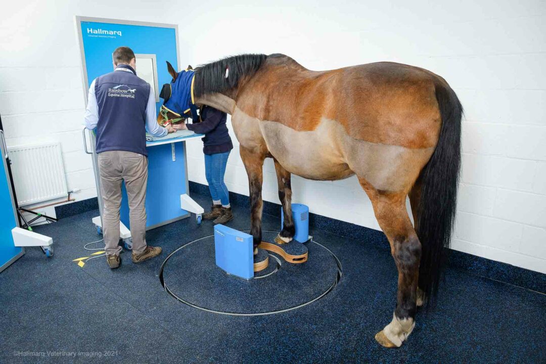
The Vision CT system features a walk-in-scan-walk-out floor-level system that guarantees easy entry and safe exit for standing sedated horses. This design eliminates the need for general anesthesia, reducing the risk to the patient while simplifying the diagnostic process. And, in combination with standing MRI, Vision CT’s ability to obtain detailed images of the foot, pastern, fetlock and distal cannon bone of both forelimbs and hind limbs, offers comprehensive diagnostics that are both rapid and risk-free.
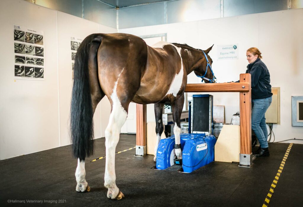

The standing MRI and CT systems complement each other by providing detailed images of bone and soft tissue – creating a complete picture of the distal limb’s health. This is invaluable for pre-operative planning, allowing veterinarians to pinpoint the precise location of pathology with unprecedented accuracy. Common applications include the diagnosis of non-displaced fractures, keratomas, pedal bone osteitis and more, which showcase the system’s versatility and effectiveness.
Research and Real-World Impact
The adoption of combination scanning at centers such as Dick Vet is not just a milestone in veterinary diagnostics, it also paves the way for in-depth research into the efficacy of these technologies. A three-year research project led by equine specialists at the facility will assess the comparative advantages of CT and MRI for detecting various distal limb problems. This research is expected to provide valuable insights to help further refine diagnostic strategies and treatment options for equine patients globally.
As veterinary medicine continues to advance, the integration of combination scanning with Vision CT and standing MRI represents a big step towards safer, more accurate and efficient equine diagnostics. With its potential to transform how lameness and foot issues are diagnosed and treated, this approach highlights the vital importance placed on innovation to ensure continuing equine health and well-being.
CT and MRI Case Study: DDFT Marginal Tear
Building on the impressive capabilities of combination scanning with Vision CT and MRI, a recent case study from Donnington Grove Equine Vets illustrates the practical application and benefits of using these technologies in diagnosing complex equine injuries. The case involved an 11-year-old Warmblood gelding presenting with acute lameness and swelling, which led to a diagnostic challenge addressed through the use of combination scanning with Vision CT and standing MRI. This case illustrates the use of dual-modality (Vision CT and Standing Equine MRI) 3D imaging to provide a pre-operative diagnosis of a marginal deep digital flexor tendon injury.
Dr Sarah Taylor from The Royal (Dick) School of Veterinary Studies, emphasized the difficulty in diagnozing marginal deep digital flexor tendon (DDFT) injuries within the digital flexor tendon sheath using conventional methods. The dual modality approach, however, facilitated a precise pre-operative diagnosis.
MRI and CT Findings: a Comparative Insight
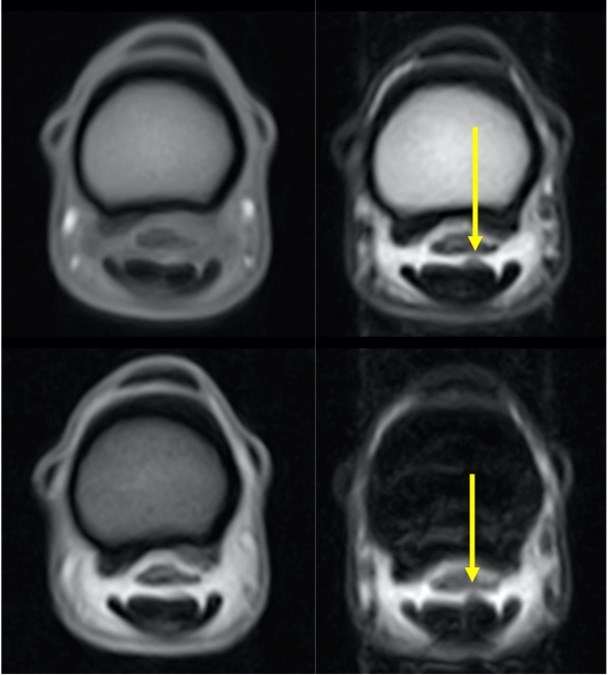
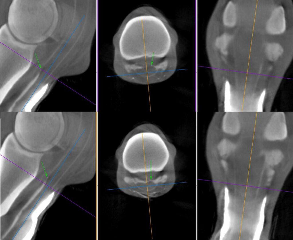
The MRI revealed abnormal curvature/enlargement of the lateral lobe of the deep digital flexor tendon, indicating the presence of granuloma or torn tendon fibres. This was complemented by CT findings that showed dorsal displacement of contrast material–– also confirming the presence of granuloma or torn tendon fibres, which was difficult to ascertain through traditional diagnostic approaches.
The Significance of Combining CT and Standing MRI
This case is a good example of the enhanced diagnostic capabilities demonstrated by the integration of Vision CT and MRI. As Dr Taylor pointed out:
“Combining these advanced techniques has the potential to improve diagnostic sensitivity and specificity for these difficult injuries within the equine digital flexor tendon sheath.”
Dr. Sarah Taylor, (BVM&S MSc PhD Cert ES(Orth) DipECVS DipECVSMR MRCVS) Senior Lecturer in Equine Orthopaedics, The Royal (Dick) School of Veterinary Studies, The University of Edinburgh
This holistic imaging approach not only pinpoints the pathology but also delineates the extent of the injury, guiding more effective treatment planning.
Operational and Clinical Advantages
The operational benefits of the standing CT system, such as avoiding general anesthesia and minimizing radiation exposure, combined with the detailed soft tissue contrast provided by MRI, present an impressive and comprehensive diagnostic tool. This approach reduces patient risk, enhances workflow efficiency and provides veterinarians with a previously unattainable level of diagnostic detail.
In Conclusion
The integration of Vision CT and MRI represents a significant advance in veterinary diagnostics – offering a more nuanced and comprehensive understanding of equine injuries. The case study is a testament to the potential of these technologies to transform equine healthcare. And as the veterinary community continues to embrace these innovations, the future of equine diagnostics looks bright––promising better care and outcomes for equine patients worldwide.
INTERESTED IN VISIONARY VETERINARY IMAGING?