In the quest for excellence in equine healthcare, the approach to diagnosing lameness has been continually and meticulously refined over the years. Today, the combined use of standing MRI (sMRI) and Vision CT scanning sits at the pinnacle of these advancements – redefining the gold standard in lameness diagnosis. We explore the synergistic power of these modalities, shedding light on the transformation they bring to equine diagnostics.
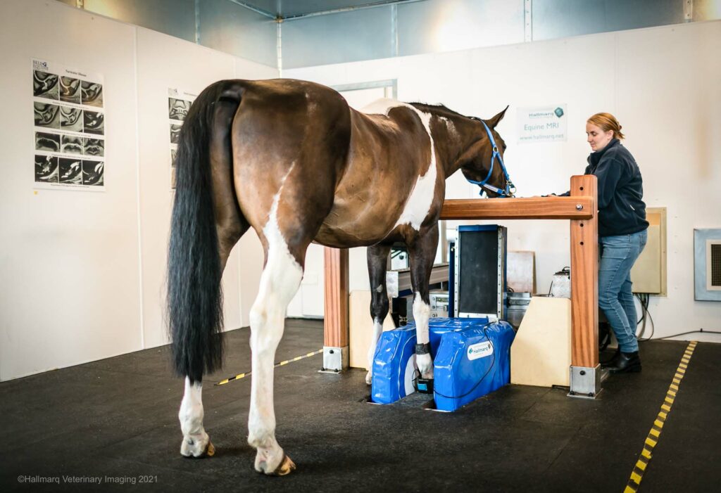
The Value of Standing MRI
The introduction of sMRI into veterinary practice marked a significant leap forward, especially when identifying soft tissue pathologies. Constituting around 70% of the cases encountered in clinical practice by Hallmarq customers, its precision in imaging soft tissues has made MRI the go-to modality, setting a benchmark in diagnosing equine lameness.
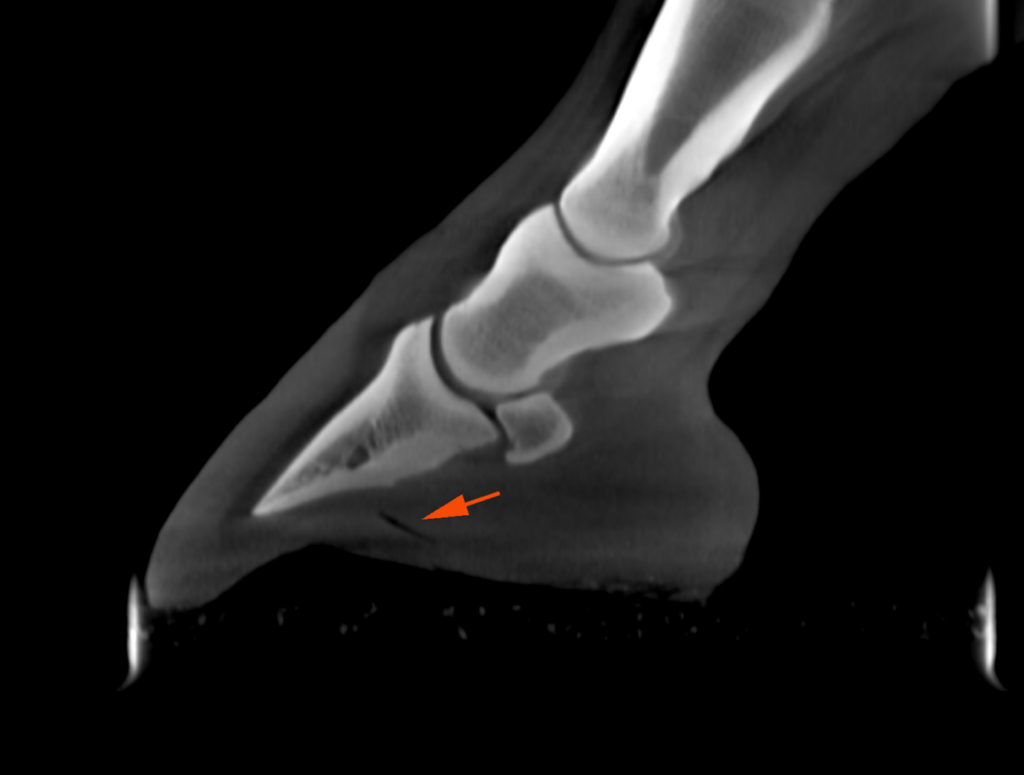
However, the diagnostic landscape is ever-evolving. While sMRI excels in soft tissue imaging, certain bony pathologies can elude it. This is where Vision CT comes in! As a complementary modality to sMRI it helps bring to light conditions like osteoarthritis, ossification of collateral cartilages, and fractures that may not be as apparent on MRI scans.
The Power of Combination Scanning
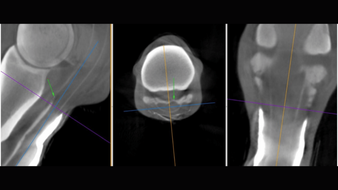
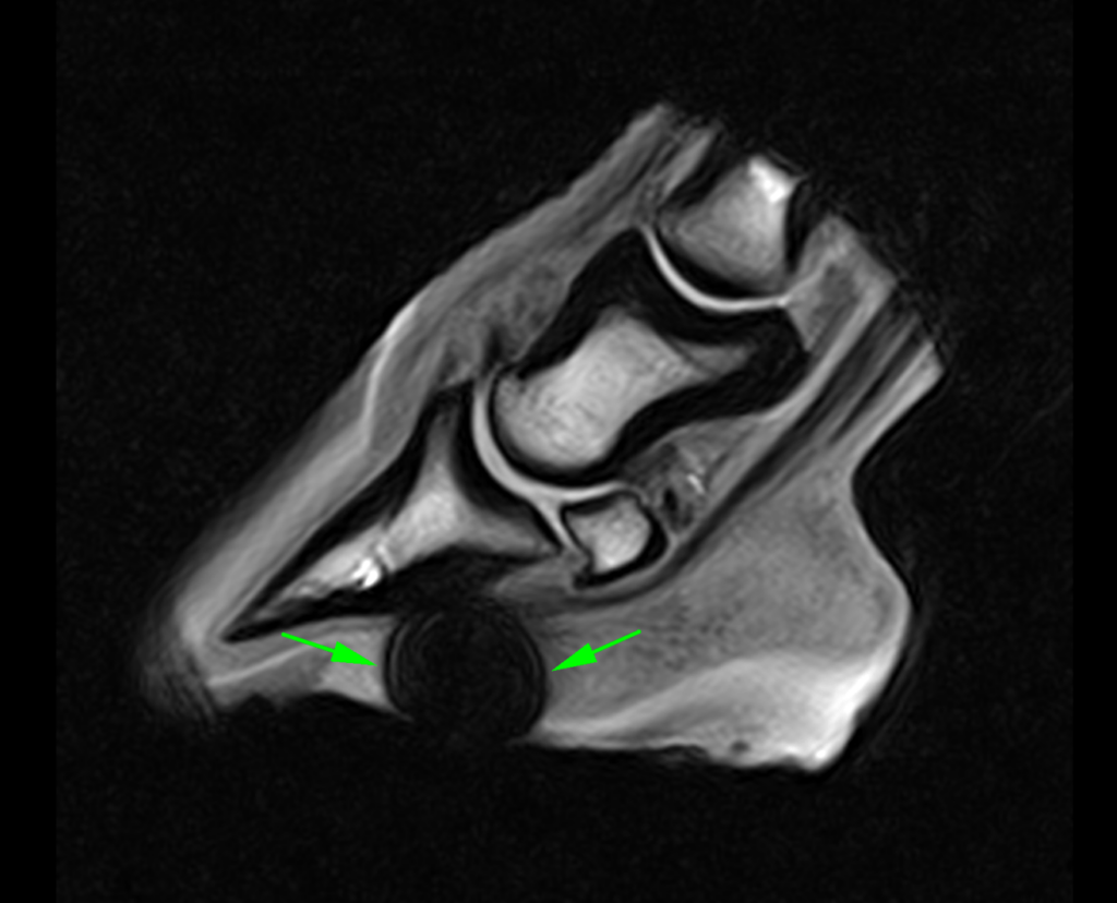
The rationale for integrating Vision CT with sMRI stems from a simple premise: more information leads to better decisions. This combination scanning approach presents a comprehensive view of the equine distal limb, marrying the detailed soft tissue imaging of sMRI – which provides the physiological data needed – with the exceptional bony detail provided by Vision CT to give additional structural data. A recent case study illustrates the power of complementary imaging when diagnosing a horse suffering from an acute penetrating foot trauma:
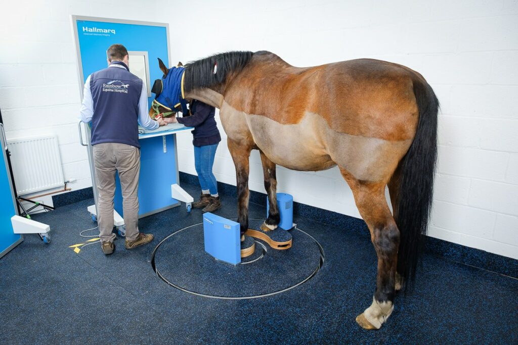
Enhancing Workflow Efficiency
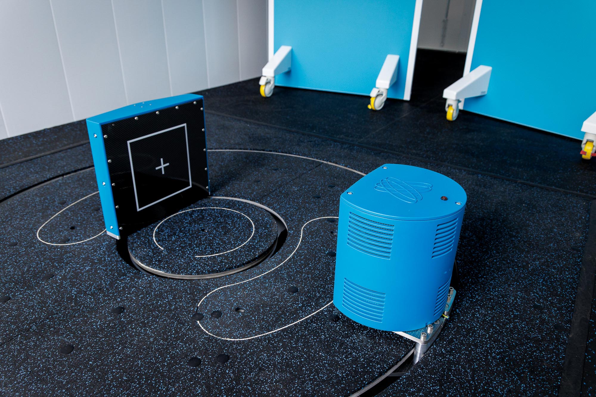
Capable of delivering 900 images in mere minutes, Vision CT offers a level of efficiency and detail far surpassing traditional radiographs. Multiplanar reconstruction allows veterinarians to view structures from any angle, significantly enriching the diagnostic process. What’s more, the radiation exposure for handlers from a single Vision CT scan is less than that from five digital radiography exposures, which speaks to its safety and efficiency.
Financial Viability and Clinical Excellence
For practices already offering sMRI, adding Vision CT can be financially viable and clinically invaluable. With flexible installation options, Vision CT’s low running costs and outpatient procedure model make it an accessible addition. Its efficiency and reduced need for anesthesia align to minimize patient risk and practice downtime.

The Incremental Cost Conundrum
Understandably, at first glance, the financial cost of adopting combination scanning might be a cause for concern or even a deterrent. However, Hallmarq has devised a cost-effective pricing model that ensures that the addition of Vision CT remains competitive for its sMRI customers. The incremental cost to the horse owner for combination scans is minimal, making it an attractive option for practices that provide top-tier diagnostic services.
Scientific Backing and Real-World Application
The clinical efficacy of combination scanning is not just anecdotal; recent studies [1] have attested to the complementary nature of CT and MRI. Research has highlighted how each modality contributes unique diagnostic data, with combination scanning offering the most comprehensive evaluation of bone and joint pathologies. Such findings are instrumental in guiding surgical planning and therapeutic decision-making, further supporting the transformative potential of this approach.
“In conclusion, all modalities were able to identify fissures with sensitivity higher in CT and specificity higher in MRI.”
Animals 2023, 13(18), 2912; https://doi.org/10.3390/ani13182912
As more practices adopt combination scanning with standing equine MRI and Vision CT, the landscape of equine lameness diagnostics is set to evolve dramatically. This integration signifies a leap forward in diagnostic precision and embodies the collective ambition for a future where every horse benefits from the highest standard of care.
The clinical benefits of this innovative approach extend beyond diagnostics into strategic therapeutic planning. The insights offered by the combination of MRI and CT scans enable veterinarians to tailor treatment strategies with a level of precision previously unattainable. The detailed images support a deeper understanding of the extent and nature of injuries, facilitating targeted interventions that can significantly enhance recovery outcomes.
The practical application and benefits of combination scanning in equine care have been echoed by professionals in the field. Elisabetta Giorio from Donnington Grove Equine Vets commented that:
“The combination of MRI and CT was a useful tool to have and helped with surgical planning and decision-making.”
Elisabetta Giorio, Donnington Grove Equine Vets
Positive, affirming responses like hers have been commonplace amongst other veterinary professionals worldwide.
A Step Towards Comprehensive Care
Providing a more definitive diagnosis by combination scanning with Vision CT and MRI paves the way for comprehensive care. This dual-modality approach ensures that no aspect of the equine distal limb’s condition is overlooked. It bridges the diagnostic gap, ensuring that both soft tissue and bony pathologies are thoroughly evaluated, offering veterinarians a complete understanding of the patient’s condition.
Empowering Practices With Innovative Solutions
The introduction of combination scanning represents a significant advancement in veterinary diagnostics, empowering practices with innovative solutions that align with the evolving expectations of horse owners. The incremental cost of adding Vision CT to existing MRI services is offset by the substantial clinical and operational benefits, making it a cost-effective option for practices aiming to elevate their standard of care.
Navigating the Future of Equine Diagnostics
As veterinary medicine continues to evolve, integrating advanced imaging techniques such as MRI and CT scanning will play a pivotal role in shaping the future of equine diagnostics. The combined strength of these modalities offers a promising path toward more accurate, efficient, and comprehensive diagnostic capabilities. It signifies a commitment to employing the best available technology that benefits equine patients and their owners.
Conclusion
The synergy of standing equine MRI and Vision CT in combination scanning redefines the gold standard in diagnosing lameness and marks an important achievement in veterinary medicine. This integrated approach enhances diagnostic accuracy and streamlines workflows to support better-informed clinical decisions. As we move forward, the continued adoption and integration of these advanced imaging modalities will undoubtedly contribute to the ongoing advancement of equine healthcare, ensuring that our equine patients receive the highest quality care possible.
The journey towards integrating combination scanning into veterinary practices is not without its challenges. However, the potential benefits for equine health and welfare are immense. As more veterinary practices embrace this technology, the future of equine diagnostics looks brighter than ever – promising a new standard of care that is both comprehensive and compassionate.
[1] Lin S-T, Foote AK, Bolas NM, Peter VG, Pokora R, Patrick H, Sargan DR, Murray RC. Three-Dimensional Imaging and Histopathological Features of Third Metacarpal/Tarsal Parasagittal Groove and Proximal Phalanx Sagittal Groove Fissures in Thoroughbred Horses. Animals. 2023; 13(18):2912. https://doi.org/10.3390/ani13182912
INTERESTED IN VISIONARY VETERINARY IMAGING?