

Technology
How it works
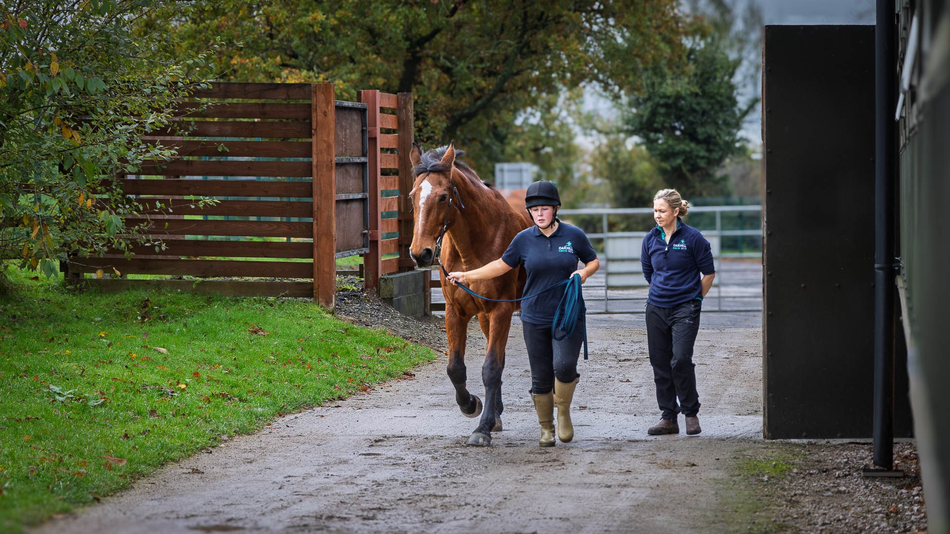
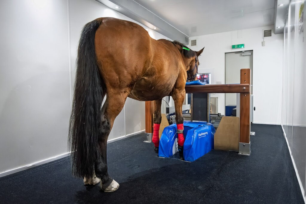
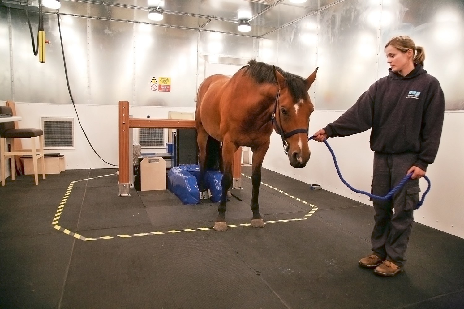
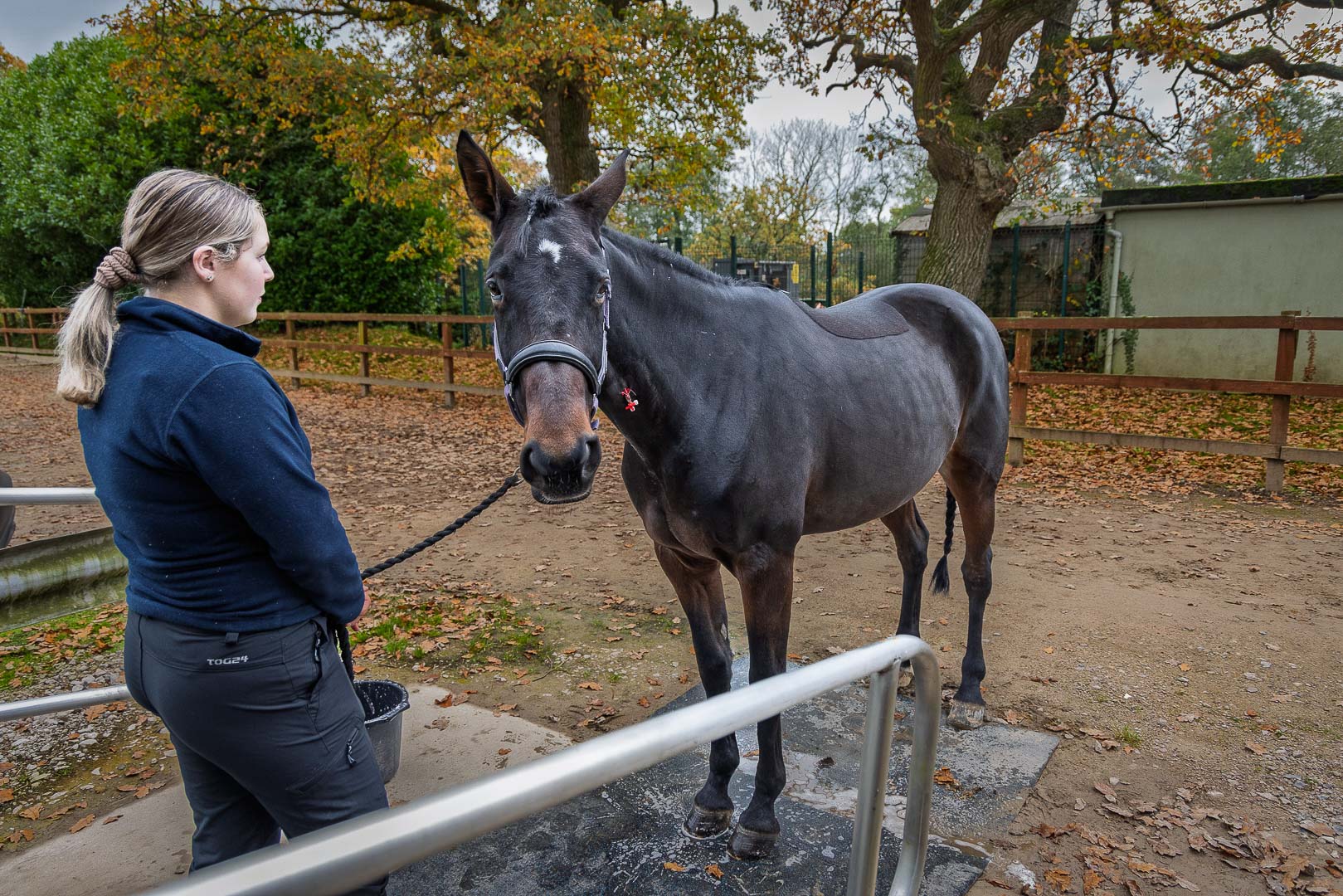
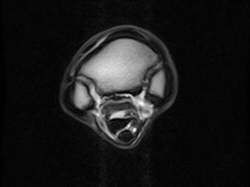
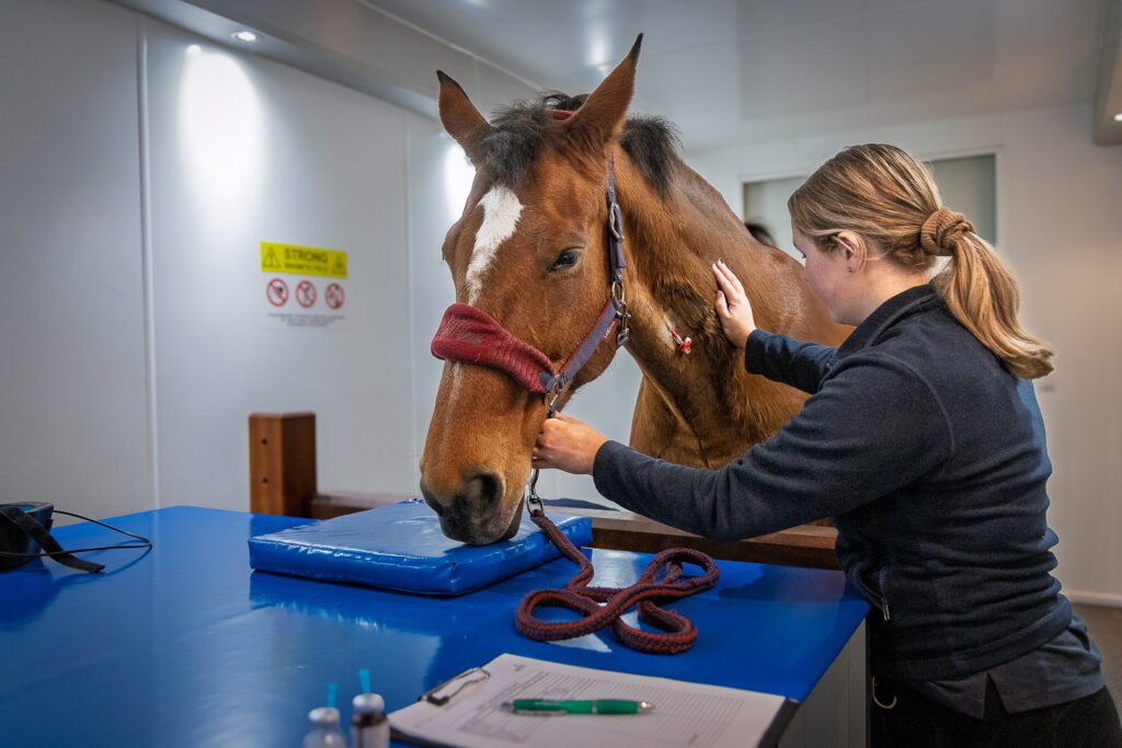
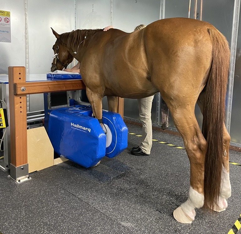
Standing Equine MRI is a low-field procedure performed on the standing, sedated horse. This unique modality visualises slices through tissue – quickly and with precision. Diagnostic in 90% of lameness cases* it can detect both bone and soft tissue abnormalities where other modalities may fail.
Widely considered the gold standard** for imaging the foot of the standing horse, your practice is more efficient and your workload more streamlined with just two cases per week.
Details
Clinical uses
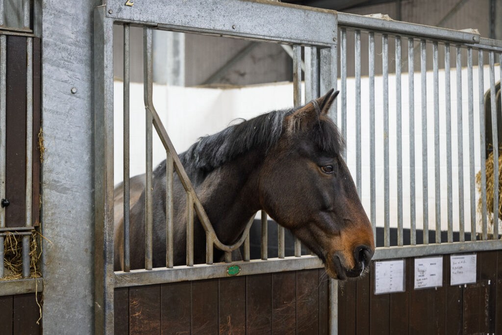
Distal limb
Diagnosis of bony and soft tissue disease when more conventional imaging techniques are negative, unclear or access is difficult
Navicular disease
MRI is the only imaging modality to provide an accurate diagnosis of this multifactorial disease, allowing for targeted treatment
See more
MRI identifies subtle lesions such as bone inflammation and early onset degenerative joint disease earlier than ultrasound and radiography
Aids surgical planning
When additional assessment to radiography is necessary (penetrating injury, fracture), standing MRI aids surgical planning without GA
Racehorses in training
With an increased risk of fracture, detecting early indicators enables modification of training, preventing catastrophic injury
Rehabilitation guidance
Tailored treatment plans and monitoring progress optimises rehabilitation. Time taken for the patient’s safe return to work is minimised
Why MRI?
Features & benefits
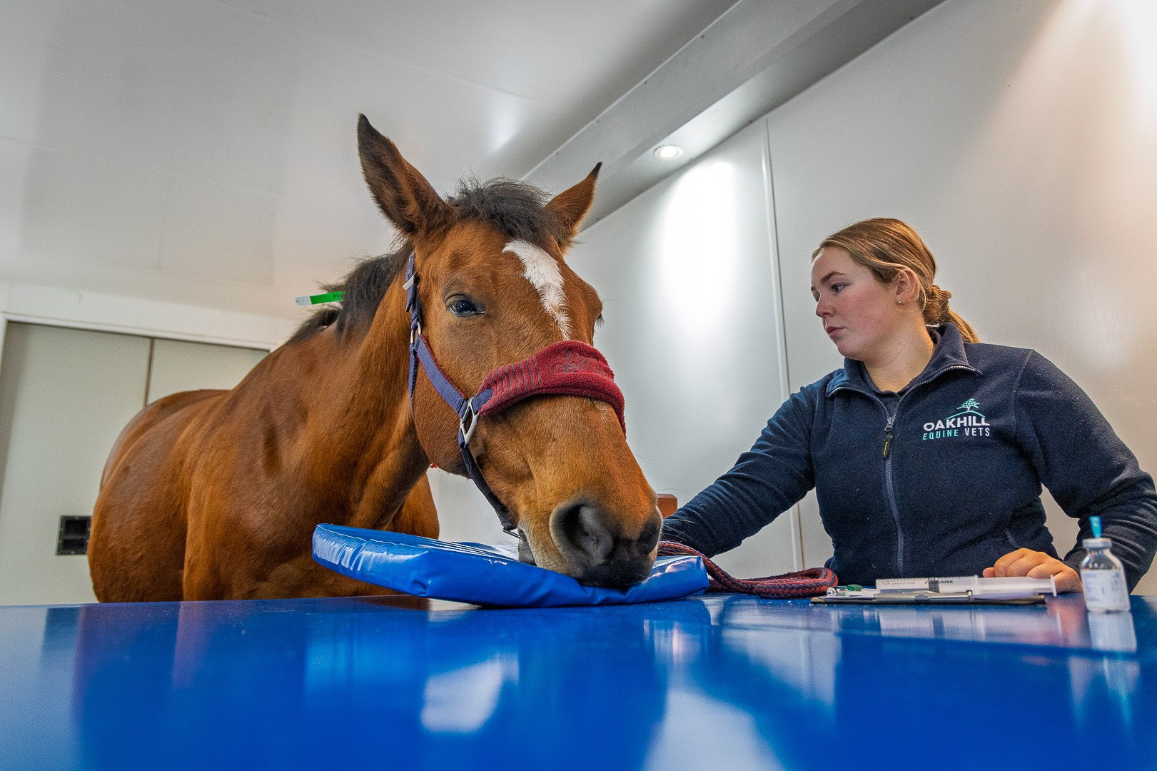
MRI without anaesthesia
No risk to the horse and a simplified workflow for your practice staff
Value add for your practice
Flexible install options & unique business models help deliver an ROI in 2 cases a week
Seamless integration
Images can be read with industry standard PACS & DICOM software
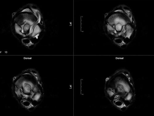
iNav Motion Correction
Reduces image artefacts caused by patient sway. Faster scan times mean reduced costs
From the ground up
Deliver the gold standard in diagnostic imaging from the hoof to the carpus or tarsus
Q-Care Cover
Outstanding remote support and a wealth of expertise to support you and your MRI
White Paper
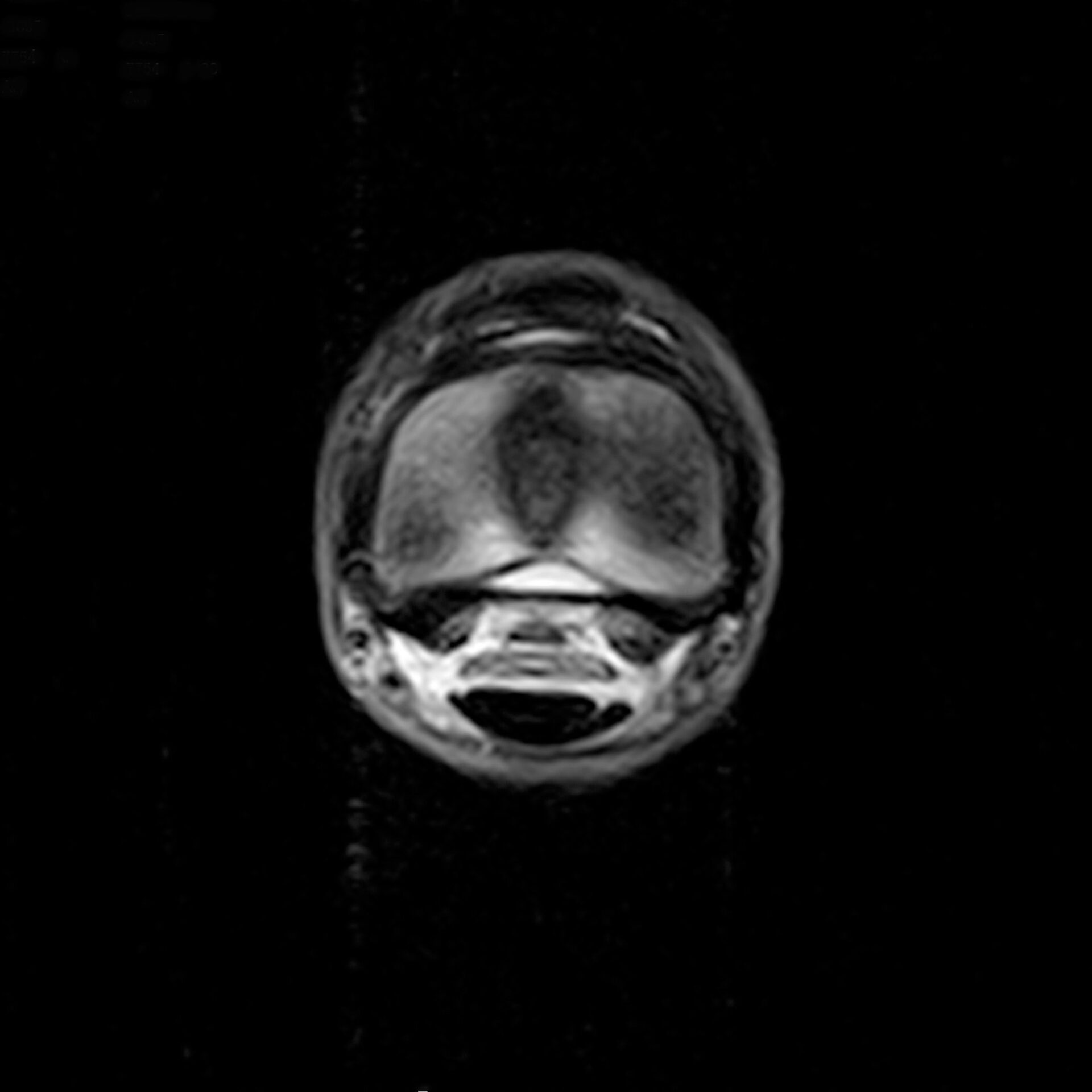
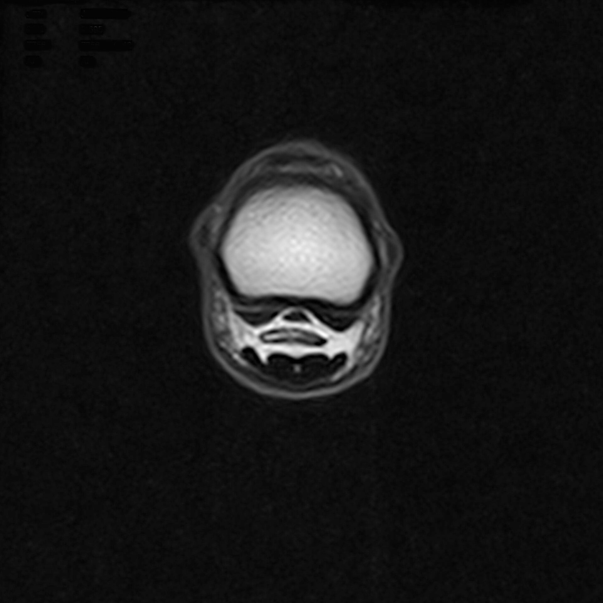
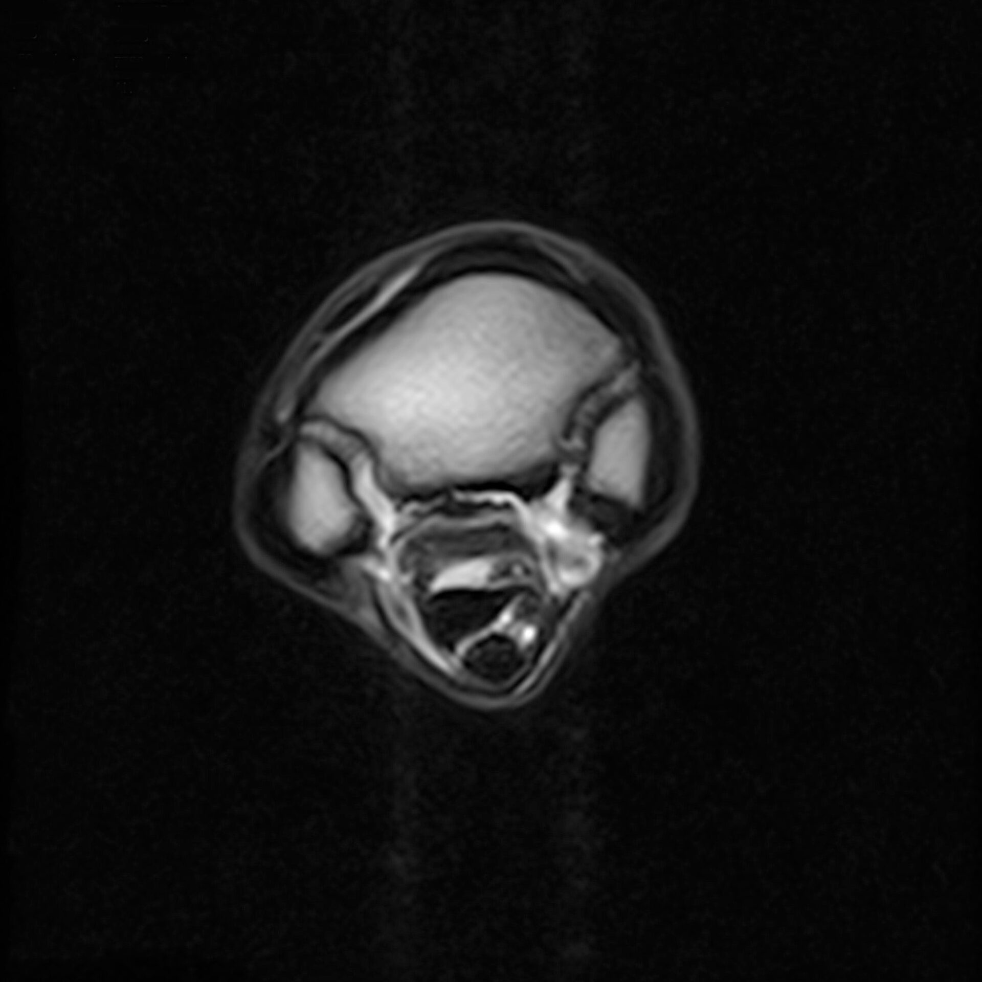
Standing Equine MRI
This paper is intended as a tool to explore the growing importance of standing equine MRI (sMRI) as a diagnostic tool for equine lameness. By highlighting the advantages of sMRI over traditional imaging methods, this paper demonstrates how integrating sMRI can add value to equine veterinary practice by enhancing diagnostic accuracy, reducing costs, and elevating the veterinary practice’s reputation and profitability.
Detailed imaging
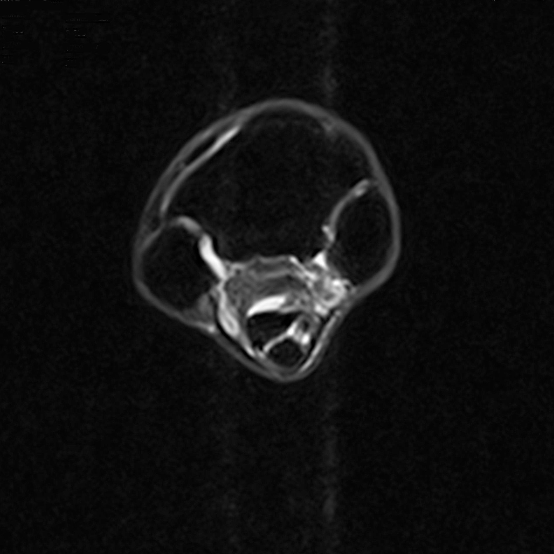
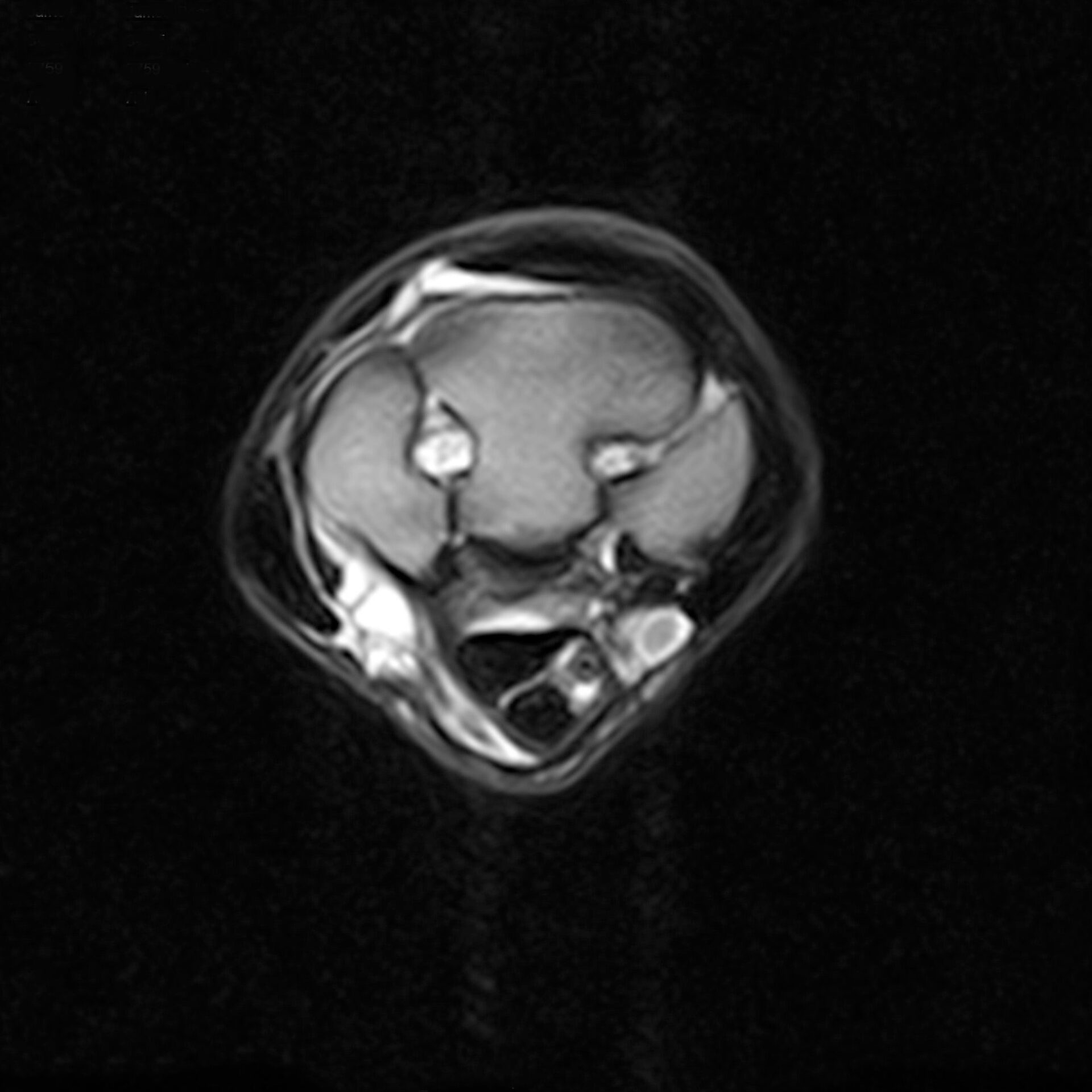
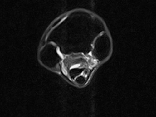
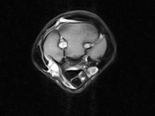
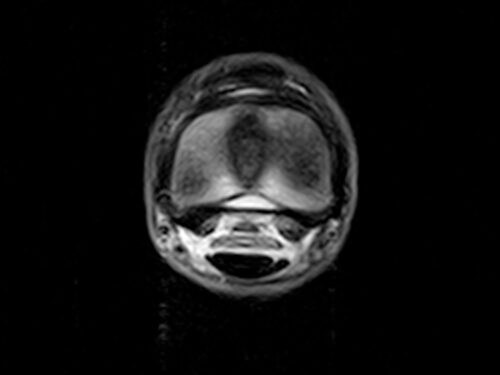
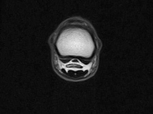
Clinical gallery
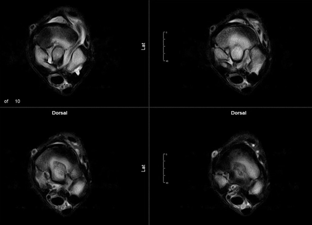
Standing Equine MRI captures superb images for a more accurate diagnosis. Now with iNAV – the latest enhancement to our unique motion correction software – you get truly transformational results.
- iNAV’s ground-breaking technology further reduces image artefacts caused by patient movement
- iNAV is particularly beneficial for scanning anything above the foot
- iNAV transforms images of the proximal suspensory, carpus, tarsus, and fetlock regions
- iNAV preserves the detail and clarity required for accurate assessments
- iNAV comes as standard for all our customers and at no extra cost
Installation options
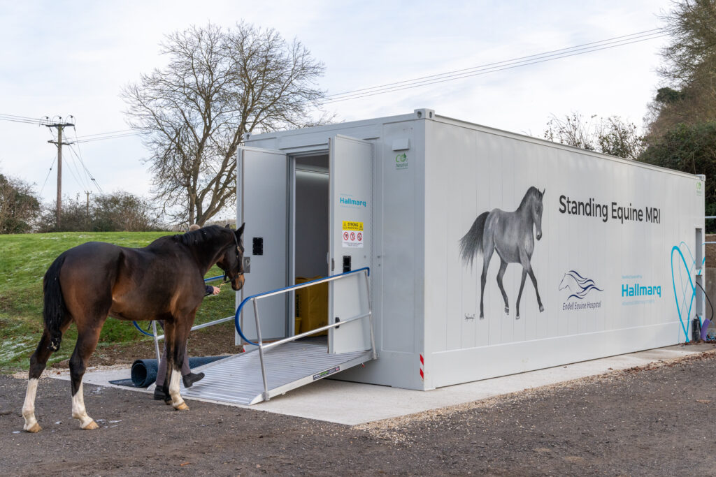
Room or modular
We appreciate that space is at a premium. That in mind, our Modular Room offers cost-effective MRI ready-to-go. Dropped into place externally to your main building, quick and easy setup means you can start scanning from day one. Alternatively, we can install a traditional room set-up
I must thank the whole Hallmarq team for your help and development of such a useful and effective tool”
Matteo Toniato DVM Clinica Equina San Biagio
The team at Hallmarq continue to find innovative approaches to improve motion correction and reduce slice thickness. When I use their images at International conferences, I am repeatedly asked if they were taken with a high field magnet.”
Sarah Taylor BVM&S MSc PhD Cert ES(Orth) DipECVS DipECVSMR MRCVS The University of Edinburgh. The Royal (Dick) School of Veterinary Studies
Hallmarq has been a great partner in providing our MRI services. The training provided was well organised and sponsorship of our MRI CE program provided to referring veterinarians was a great success. Thank you Hallmarq for your continued support.”
Dr. Bill Hay DVM DACVS Tryon Equine Hospital Columbus, NC
Hallmarq’s MRI has revolutionised the way foot lameness has been diagnosed in thousands of cases over the last 15 years. Excellent image quality facilitates accurate diagnoses, appropriate treatment selection and formulation of a prognosis.”
Sarah Taylor BVM&S MSc PhD Cert ES(Orth) DipECVS DipECVSMR MRCVS The University of Edinburgh. The Royal (Dick) School of Veterinary Studies
The team at Hallmarq provide superb customer support, are friendly, approachable and always happy to help. This enables the user to make the most of the system and optimise image quality.”
Dr Rhiannon Morgan BSc BVSc CertAVP MVetMed(Hons) FHEA PhD DipECVDI MRCVS IVC Evidensia
Advanced diagnostic imaging techniques have become increasingly within reach for countless horses over the past 15 years and Hallmarq has played a large part in this rapid and exciting evolution within the equine industry.”
M.A. Smith MA VetMB Cert ES DipECVS AECVDI MRCVS Image Equine

Accurate
Diagnostic in over 90% of cases, Standing Equine MRI can accurately identify the specific cause of lameness and the degree of pathology which leads to a full understanding of the injury. This means than an optimal treatment plan can be initiated immediately with confidence.
Convenient
Bring all the benefits of Standing Equine MRI to your practice without the complications of general anaesthesia. The horse remains standing throughout and, in most cases, can be seen as a day patient.
Affordable
Low upfront costs include the option to include a modular solution or a purpose built room. A unique pay-as-you-go model means that, with approximately 2 cases per week, your Standing Equine MRI soon pays for itself.
FAQ’s
Frequently asked questions
MRI is unparalleled in providing images of both soft and bony tissues. Distinguishing water from fat, it highlights areas of pathology such as inflammation and bruising, in a way that radiography, CT, ultrasound or nuclear scintigraphy just can’t do. By imaging the region of interest in slices orientated in any 3D plane, a lesion can be visualised without superimposition of adjacent structures. Multiple views allow you to appreciate the full extent of the injury.
If you’re a referring vet looking to have better conversations with your horse owners, take a look at our educational resource – Talk Lameness. Aimed at answering questions around lameness, which can be confusing, it’s designed to provide reassurance with what can often be a difficult topic to pin, and to encourage conversations between vet and owner.
Find a system
Our systems are available at some of the world’s leading veterinary facilities around the world. To find a Hallmarq MRI or CT machine near you, click below.
Find a siteTechnical
Specification
In planning for the installation of your Hallmarq Standing Equine MRI system, the high-level technical specifications below should be taken into consideration. Please consult the Site Planning Guide for additional details.
| Magnet | |
|---|---|
| Magnet field strength | 0.27 Tesla (+/- 0.02 Tesla) |
| Pole gap | 220mm (+/- 10mm) |
| Imaging volume | 140mm DSV |
| Homogeneity | <110 PPM (peak to peak) measured over a 150 mm DSV |
| Position | 3-dimensional magnet positioning system |
| Rotation | 90-degree magnet rotation |
| Movement device | Vertical and in/out motorised movement with manual control. |
| Manual left/right movement via side wheel. |
| Gradients | |
|---|---|
| Three orthogonal imaging gradients, plus one Z2 channel | |
| Maximum imaging gradient strength: 25 mT/m | |
| Maximum imaging slew rate: 75 mT/m/ms |
| RF Coils | |
|---|---|
| 4 x coils of different sizes for imaging the foot and lower limb |
| Electronics | |
|---|---|
| Spectrometer | Digital RF unit running at 11 MHz (+/- 1 MHz) |
| RF and gradient amplifiers | |
| Computer | Multi-core Intel processor with dual solid-state drives |
| Windows 10 LTSC operating system | |
| Pulse sequence library with advanced motion correction | |
| 24″ wide screen monitor, mouse and keyboard |
| Software | |
|---|---|
| MRI software suite | |
| Pulse sequence library with advanced motion correction | |
| Remote access and management via LogMeIn |
Q-Care
Supporting your success
Providing high-quality images is just the beginning. With an unrivalled programme designed to add value to your practice, we’re here to help optimise your imaging service – from initial enquiry through to installation and beyond.

* (Byrne, Marshall, and Voute, 2020)
** (Gutierrez-Nibeyro et al. 2012, Sherlock et al. 2007, Mair et al. 2005, Olive et al. 2009)

