

Why are carpus scans increasing?
The growing adoption of carpus and tarsus MRI scans among equine vets has partly been influenced by software updates to the Hallmarq system. These improvements were developed to combat image problems resulting from motion of the patient during scanning. The swaying movement of horses being scanned becomes more problematic higher up the leg as the increased motion is more detrimental to image quality. Software developments by Hallmarq can now correct for a greater degree of motion and even compensate for change in shape of a joint during the scan.
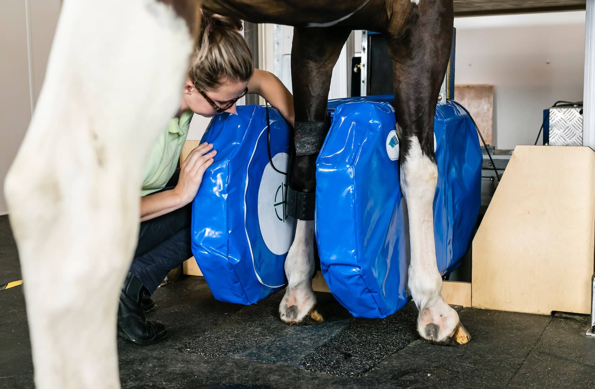
The primary strength of the Hallmarq MRI is that horses can be scanned while standing. However, the magnet can be rotated by 90° to scan a horse under general anaesthetic if required. This is sometimes used for scans of the hock as some horses object to having the magnet positioned between the hind legs.
What makes MRI different?
If an injury is suspected in the leg then X-rays and ultrasound are often the first port of call.
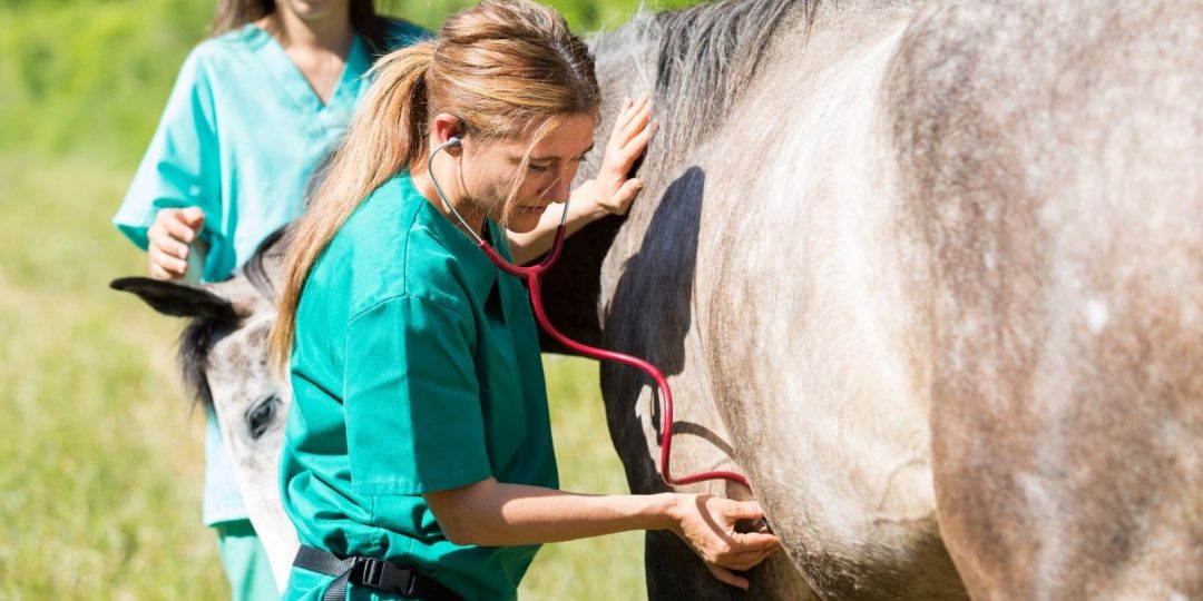
Ultrasound is widely used for imaging soft tissues in both human and veterinary medicine. It’s a relatively cheap technique, but the grainy images can be difficult to interpret and rely a lot on the skill of the operator. Ultrasound waves will not pass through bone, so the technique is particularly difficult to use around the bones of the carpus or the splint bones in the upper leg. However, as studies have shown that stem cell therapy is more effective at treating tendon lesions when it is performed soon after the injury, a quick diagnosis will help to increase the chance of a good recovery. MRI gives more detailed information on the condition of the leg’s tendons and ligaments, and seeing slices taken in multiple directions allow complete evaluation of the area.
X-ray is the primary method of assessing bone condition. Again it is relatively inexpensive and can often be used without bringing the horse into the clinic. Its main limitation is that the two-dimensional images produced show shadows of overlying bones. This makes it hard to completely evaluate the complicated structure of the carpus and tarsus where multiple bones overlap. Occasionally, a horse might be diagnosed with a fracture via X-ray, but not respond to treatment as expected. In these cases an MRI might reveal that nearby soft tissue structures have also been affected and require treatment.
Certain fractures or pre-fracture bone changes can also be missed on X-ray. MRI can detect fluid in bone that is often the precursor of a fracture. This is particularly common in racehorses since their carpal and fetlock joints are under a lot of strain during fast work. Spotting the damage early allows the owner to change the horse’s management and avoid potential injury.