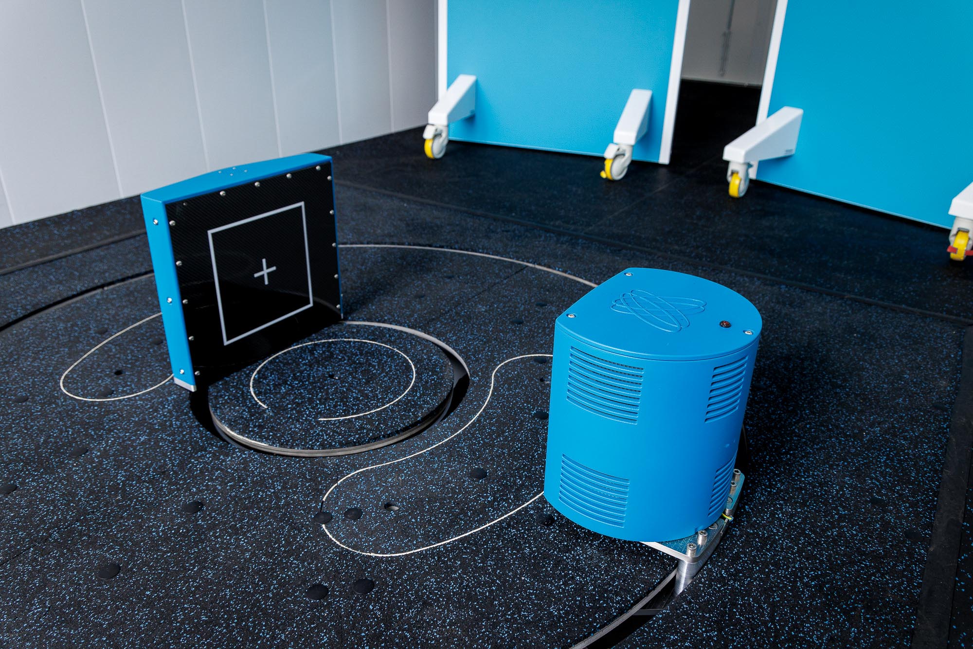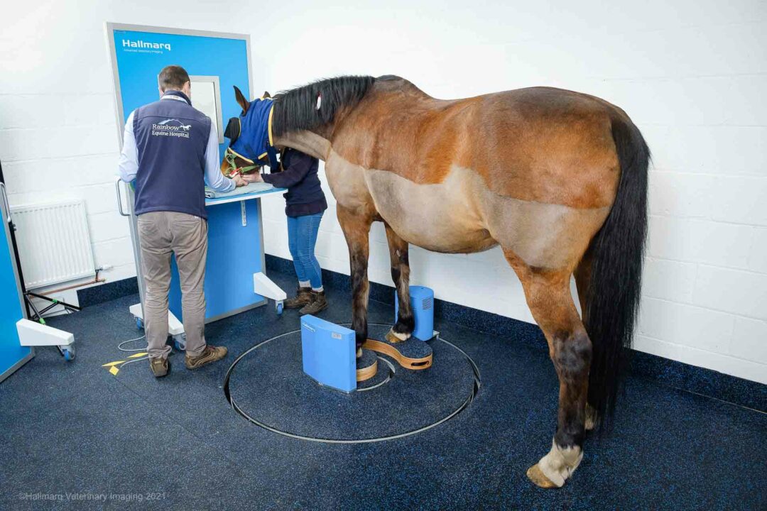
Designed to bring fast 3D imaging to every equine practice, Hallmarq’s Vision CT offers a unique walk-in-scan-walk-out system to provide safe, effective, and affordable advanced imaging for every equine practice. It’s the next step up from DR and is proving particularly effective when used in combination with our Standing Equine MRI.
A growing body of published, peer-reviewed clinical evidence is now widely available comparing Cone Beam CT (CBCT), Fan Beam CT (FBCT), and Magnetic Resonance Imaging (MRI). We take the opportunity to round up the papers for review:
Sagittal and parasagittal groove fissures
This study sheds light on the potential applications of CBCT in veterinary imaging.
Although no significant difference was observed between Cone-Beam CT (CBCT) and Fan-Beam CT (FBCT), it is important to note that the study was conducted using cadaveric limbs. Motion artifact, which can diminish image quality, is not accounted for in CBCT and MRI, thus rendering FBCT the most sensitive modality, since horses are under general anaesthesia.
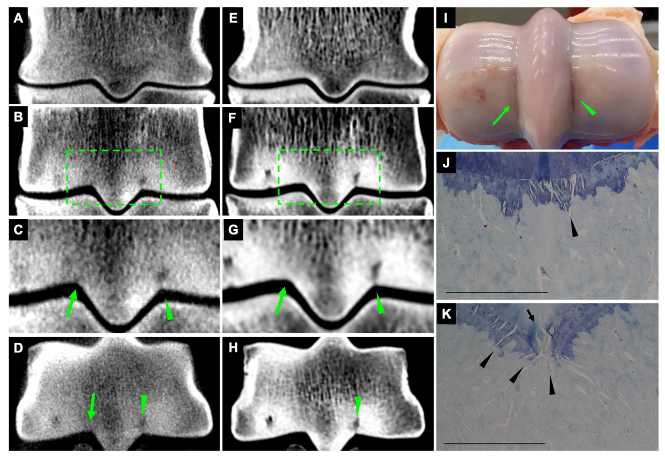
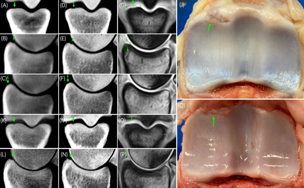
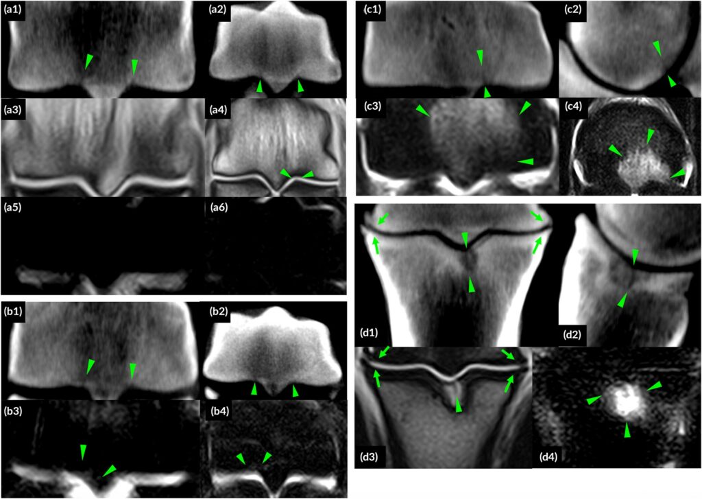
Palmar osteochondral disease
Palmar/plantar osteochondral disease (POD) of the metacarpal/tarsal condyles is a common pathological finding in racehorses. This cross-sectional study compares diagnoses, imaging details, and measurements of POD lesions between Cone-Beam CT (CBCT), Fan-Beam CT (FBCT), and low-field magnetic resonance imaging (MRI) using macroscopic pathology as a gold standard.
Thirty-five cadaver limbs from 10 horses underwent CBCT, FBCT, MRI, and macroscopic examination. Images were examined for the presence of POD, imaging details of POD, and measurements of POD dimensions and areas.
Heterotopic mineralisation
Combining Cone-Beam CT (CBCT) and MRI can provide a more wholesome idea of the extent of the problem with everything performed on the standing horse, with no general anaesthesia required. CBCT and FBCT systems were generally superior in identifying heterotopic mineralisation than MRI. However, there were locations (mineralisation of the oblique sesamoidean ligament and ossification of the joint capsule) where CBCT failed to identify abnormalities compared to FBCT. However, CBCT is not limited by general anaesthesia.
MRI on the other hand, provided information on soft tissue pathology related to the lesions. This may be important for case management and detecting the importance of the condition as a lot of the heterotopic mineralisation is considered an incidental finding
The combined utilisation of CBCT and MRI offers a more comprehensive assessment of equine conditions, all conducted on standing horses without the necessity of general anaesthesia. It would be prudent to avoid asserting that CBCT and FBCT yield identical results. This is because such a claim does not align with the study’s findings and because the study was conducted using cadaver limbs.
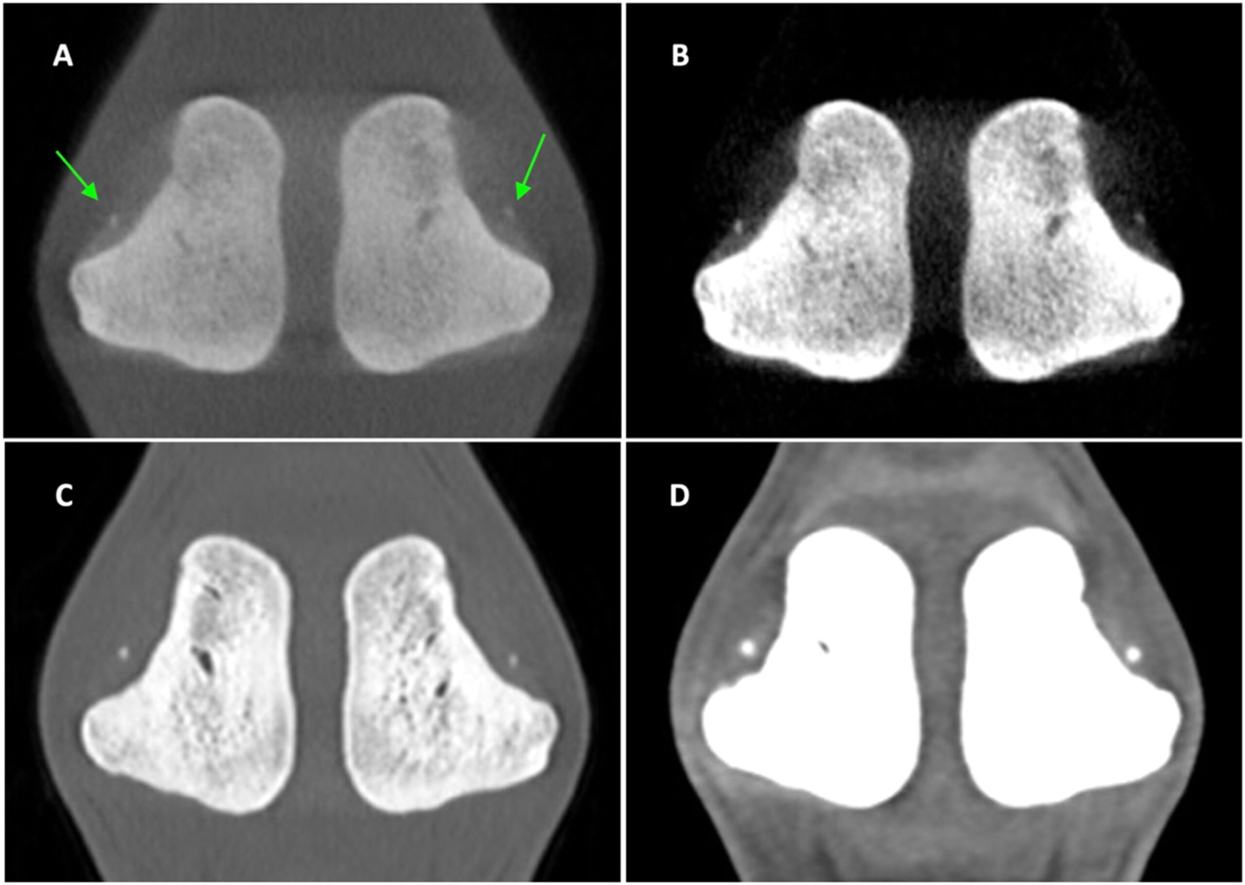
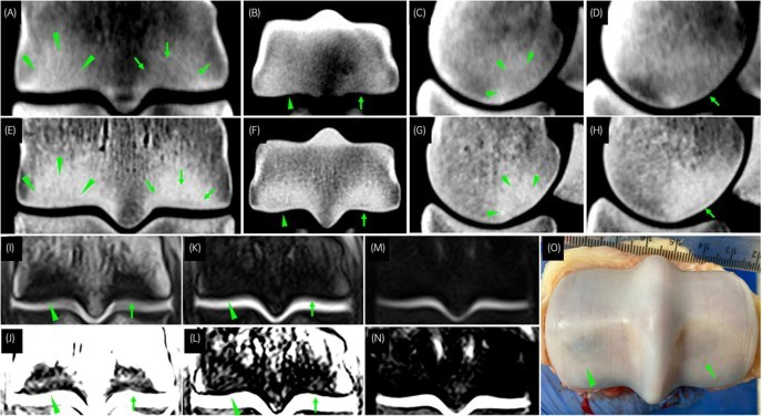
Proximal phalanx dorsoproximnal osteochondral defects
Fan-Beam CT (FBCT) exhibited superior diagnostic performance when compared to CBCT and MRI, positioning FBCT closer to the gold standard, particularly in discerning smaller-sized defects. However, CBCT demonstrated the second-highest diagnostic efficacy. When CBCT is coupled with MRI, a more comprehensive assessment of equine osteochondral conditions can be achieved, as MRI excels in evaluating the active stage of lesions. Utilizing standing imaging techniques may present a viable approach for diagnosing osteochondral defects.
Motion artefact should be regarded as a notable limitation that was not considered in this study meaning that the sensitivity of both standing modalities may decrease further. Notably, there were no discernible differences in the features of osteochondral defects between CT and low-field MRI, indicating that radiologists may not require specific training, as lesions exhibit similar appearances across these modalities.
Imaging the metacarpo (MCP)/metatarsophalangeal (MTP) region
The study compared Cone Beam CT (CBCT – Hallmarq Vision CT) and low-field magnetic resonance imaging (MRI – Hallmarq Standing Equine MRI) findings in the metacarpo (MCP)/metatarsophalangeal (MTP) region of clinical patients imaged for diagnostic purposes.
CBCT and low-field MRI contributed different diagnostic information so optimal information was obtained using both modalities. Standing CBCT showed changes in bone structure and attenuation, but was limited in detecting soft tissue injuries. Standing MRI showed increased fluid accumulation in bone and soft tissue injuries but was limited in assessing structural details of bone injuries.
See more with Vision CT
Our comprehensive case study library is compiled from clinical case studies provided by Hallmarq customer sites. It includes cases where CT is used as a stand-alone modality and several where CT is used to complement Standing Equine MRI. You can see more here >>
INTERESTED IN VISIONARY VETERINARY IMAGING?