


Chiari-like malformation is a developmental condition that particularly affects small and brachycephalic breeds such as Cavalier King Charles Spaniels, Chihuahuas, and Pugs. Hallmarq’s Small Animal Medical Director and Advisory Board Chair, Dr. Simon Platt, explores how MRI can help diagnose Chiari-like malformation in dogs, assessing its severity, and providing insights into the prognosis.
Symptoms of Chiari-like malformation in dogs
Chiari-like malformation occurs when the skull is too ‘small’ to accommodate the brain. This causes the cerebellum to be indented or impacted into, or herniate through, the foramen magnum, the opening at the base of the skull. This can lead to a range of signs, which include sporadic pain phenomena, neck sensitivity, and even neurological deficits.
The pain can be position-based causing a refusal, hesitation, difficulty, or vocalization when going up the stairs and yelping when greeting the owner. It can often be accompanied by; an unusual head posture when sleeping and when awake. The dogs can also exhibit head pain, manifested as scratching or rubbing the head or ears and sometimes yelping whilst scratching these areas. In addition, they can have an aversion to touch or grooming of the head and ears but tolerate touch or grooming elsewhere. There can be altered sleep patterns in some affected dogs, with altered activity levels and becoming more timid, anxious, or withdrawn in general.
Magnetic Resonance Imaging (MRI) has become an invaluable tool in diagnosing Chiari-like malformation in dogs, assessing its severity, and providing insights into the prognosis.
Diagnosing Chiari-like malformation (CM) with MRI
MRI is the gold standard for diagnosing Chiari-like malformation in dogs, which helps rule out other conditions that could cause similar clinical signs. Unlike X-rays or CT scans, MRI provides detailed, high-resolution images of the brain and spinal cord. MRI allows veterinarians to visualize the structural abnormalities associated with CM.
Ideally, predisposed breeds would have MRI screening from 12 months of age (before breeding) and after 5 years old (after breeding).
Key diagnostic features of Chiari malformation on MRI include (Figure 1):
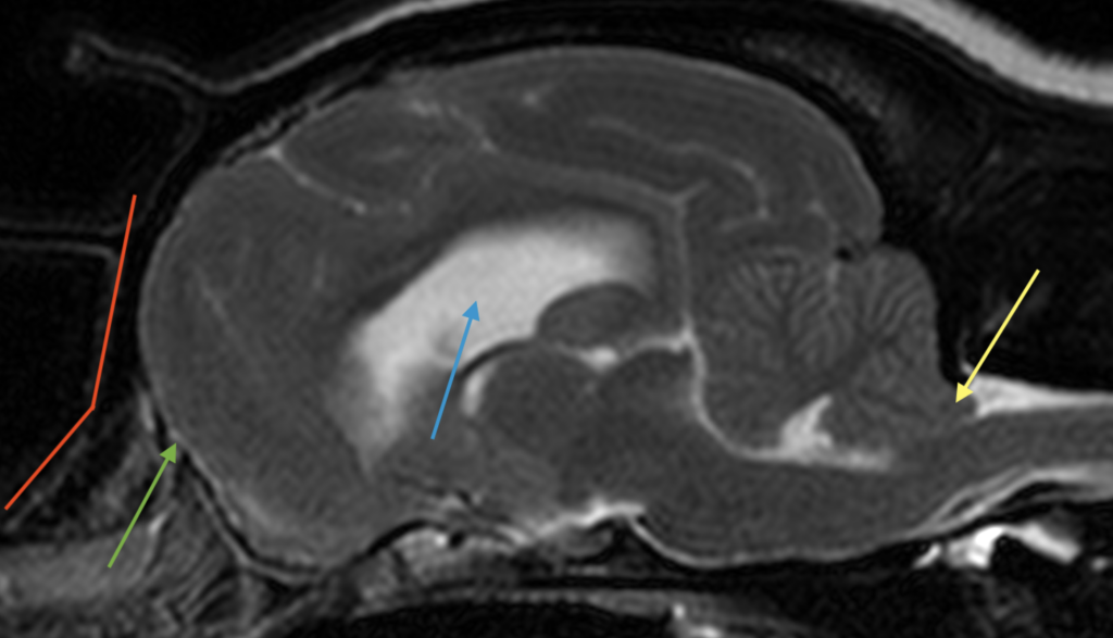
- Skull and head shape change – the frontal sinuses are smaller than normal or absent; the muzzle is short in height and length, and there is often a sharp angle between the nasal and frontal bones, with a shortened base of the skull.
- Forebrain shape change – both sides of the forebrain (cerebrum) become more round in shape, from rugby ball shaped.
- Enlarged ventricles
- Cerebellar herniation: The cerebellum is impacted into or protrudes through the foramen magnum
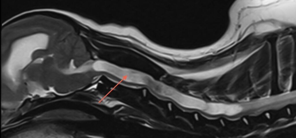
- Syringomyelia: A fluid-filled cavity (syrinx) within the spinal cord, often associated with CM, can be detected (Figure 2).
- Skull abnormalities: MRI can reveal a misshapen or undersized skull, which contributes to the condition.
Assessing the severity of Chiari-like malformation with MRI
MRI not only confirms the presence of Chiari-like malformation but also helps assess its severity. The degree of cerebellar herniation and the size of a syrinx (if present) are critical indicators of the condition’s severity. For example:
- Mild cases: Minimal cerebellar herniation and no syrinx may cause mild or intermittent symptoms.
- Severe cases: Significant herniation and a large syrinx are often associated with more pronounced neurological deficits and chronic pain.
- A grading system is provided by the British Veterinary Association
Using MRI to determine the prognosis for dogs with Chiari-like malformation
The information gathered from an MRI plays a crucial role in determining the prognosis for dogs with Chiari-like malformation. Dogs with mild herniation and no syringomyelia may have a better prognosis and respond well to conservative management, such as pain relief and anti-inflammatory medications. However, dogs with severe herniation and large syrinxes may require surgical intervention to alleviate symptoms and prevent further neurological damage if medical therapy has not adequately improved the pet’s quality of life.
In large part, the outcome of affected dogs relates to their age. Dogs that are presented with a syrinx before 3 years of age have a poorer prognosis and are more likely to develop severe weakness, which is more difficult to manage.
MRI also helps monitor disease progression. Repeat scans can track changes in the size of a syrinx or the degree of herniation, guiding treatment decisions and providing owners with a clearer understanding of their dog’s long-term outlook.
Conclusion
MRI is an essential tool in the diagnosis, severity assessment, and prognosis of Chiari-like malformation in dogs. Its ability to provide detailed images of the brain and spinal cord allows veterinarians to make informed decisions about treatment and management and rule out other conditions. For dog owners, an MRI can offer clarity and hope, enabling them to provide the best possible care for their furry companions. If your dog is showing signs of Chiari malformation, consult your veterinarian about the possibility of an MRI to ensure an accurate diagnosis and tailored treatment plan.