
Understanding Dog MRI, Cat MRI, and Small Animal MRI
Magnetic Resonance Imaging (MRI) is a non-invasive imaging technique that allows vets to get a detailed and comprehensive view of their pet’s internal structures. Dog MRI, as well as MRI for cats and other small animals, can be performed to help diagnose various conditions. This advanced technology uses powerful magnets and radio waves to create exact images without the need for surgery or exposure to harmful radiation and is an incredibly safe clinical modality.
MRI provides amazing clarity of the body’s tissues. It is the imaging tool most often recommended by veterinary neurologists and radiologists to evaluate the nervous and – sometimes – the musculoskeletal system. In contrast to CT, which is better for bone evaluation, MRI is significantly better at imaging soft tissues, such as the brain, spinal cord, intervertebral discs, tendons and ligaments, and muscles. The smallest of abnormalities can be detected with MRI that may otherwise be missed on CT and other imaging techniques. MRI also allows imaging from all three planes of the body, left-to-right, front-to-back, and top-to-bottom, without having to move the patient. This allows your vet to view the body three-dimensionally.
MRI is a critical step for getting an accurate diagnosis, particularly if your cat or dog displays signs of neurological issues like seizures, balance problems, behaviour changes, weakness, or pain. The clear, in-depth images generated by MRI allow your vets to pinpoint the exact location and severity of the issue, enabling them to formulate a precise treatment plan.
This makes MRI an invaluable tool in diagnosing complex conditions, leading to better-informed decisions and improved chances for recovery. In the case of beloved pet cat Stormy (right), MRI was integral to diagnosing feline intervertebral disc extrusion, a large slipped disc that causes compression of the spinal cord. Following surgery, Stormy has gone on to lead a full and active life.

How does MRI for pets work?
A dog MRI, cat MRI, or MRI for any other small animals is a safe and completely non-invasive procedure, meaning it does not require surgery or incisions. Instead, it relies on strong magnetic fields and radio waves to create detailed images of your pet’s internal structures. Unlike X-rays, which use harmful radiation, an MRI is particularly useful for examining soft tissues like the brain, spinal cord, muscles, and organs without causing any harm or discomfort to your pet.
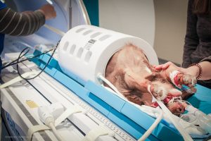
During an MRI scan, your pet is placed in a specially designed machine that houses a strong magnet. The magnetic field aligns the water molecules in your pet’s body, and radio waves are used to detect their response. This data is used to generate high-resolution images of the internal structures, helping vets detect conditions such as tumours, disc issues, or other abnormalities.
In non-emergency situations, the MRI scan typically lasts between 45 minutes to 2 hours, depending on the complexity of the area being examined. To ensure everything runs smoothly, you should plan to arrive about 30 minutes ahead of the appointment to complete any necessary paperwork and allow the veterinary team to prepare your pet for the procedure. Since pets need to remain completely still during the MRI, light anaesthesia is often required to keep them calm and relaxed throughout the scan.
Why does my dog, cat, or small animal need an MRI?
An MRI scan provides exceptionally detailed images of soft tissues, making it an invaluable tool for detecting conditions such as tumours, spinal issues, and joint problems in pets. By offering a clear and precise view of areas that are difficult to examine with other imaging techniques, MRI helps vets accurately pinpoint the source of issues affecting the brain, spinal cord, muscles, or joints.

In neurology, the clinical signs (or symptoms) exhibited by a pet depend on where the disease is located rather than what disease process is present. In other words, a brain tumour, stroke, or infection in the same area of the brain causes very similar clinical signs so the cause of your pet’s illness can’t be determined on examination alone.
As a result, advanced imaging is required in most cases. MRI allows your vet to quickly and safely provide a more accurate diagnosis of your pet’s illness, in turn, they can then more accurately treat and provide an improved quality of life for your four-legged friends. Signs and symptoms in pets that may indicate the need for an MRI might include:
- Persistent lameness or limping that doesn’t improve with rest or treatment
- Neurological changes, such as seizures, sudden balance problems, or disorientation
- Unexplained pain that doesn’t respond to conventional treatments
- Weakness in the legs, difficulty walking, or wobbly movements
- Behavioral changes, such as unusual aggression or confusion
- Sudden loss of coordination or control over bodily functions
Recognising these symptoms early is crucial. Early detection through MRI allows for a timely and accurate diagnosis, which can significantly improve the success of treatment. Whether it’s identifying a tumour, detecting spinal cord damage, or addressing joint issues, acting quickly on these signs can lead to better outcomes and a more effective treatment plan for your pet.
What conditions can MRI help diagnose in my pet?
MRI is particularly useful for examining soft tissues like the brain, spinal cord, muscles, and organs without causing any harm or discomfort to your pet. Some of those conditions are listed below in more detail here and give a good overview of the versatility of MRI in small animal veterinary practice.
Spinal cord disease
Including spinal cord neoplasia, intervertebral disc disease, wobbler’s disease and lumbosacral disease, atlanto-axial subluxation, ischemic myelopathy, nerve root compression and discospondylitis.
Brain disease
Including, but not limited to neoplasia, cerebrovascular events, hydrocephalus, and inflammatory brain disease.
Skull and surrounding soft tissues
The only modality able to accurately assess otitis externa-media-interna, including assessment of associated cranial nerves, cochlea and semi-circular canals along with the adjacent brainstem. Nasal and orbital diseases can also be clearly assessed.
Cranial and vertebral anomalies and genetic malformation screening
Assessment of congenital anomalies including vertebral malformations, Chiari-like malformation, and syringomyelia screening in predisposed breeds.
Stifle (knee) disease
Superior soft tissue imaging for assessment of cranial cruciate ligaments, meniscal injuries and muscular disease.
Shoulder disease
Superior soft tissue imaging for assessment of biceps and supraspinatus tendon pathology, joint capsule, brachial plexus disease and muscular disease.
Thoracic and abdominal disease
Complementary to ultrasound and computed tomography, soft tissue diseases affecting the lungs, liver, kidneys, spleen, and major vessels can be accurately assessed with MRI.
Epilepsy screening
Confirmation of idiopathic epilepsy requires an imaging investigation to rule out structural causes, toxicities and metabolic diseases. If an MRI of the brain is considered normal, when accompanied by a normal CSF analysis, the cause of seizures is compatible with an idiopathic disease.
Is MRI safe for my dog, cat, or small animal?
An important benefit of MRI scans is that they do not use ionising radiation, unlike X-rays or CT scans. This makes MRIs a safer option for your cat or dog, as they avoid the potential risks associated with radiation exposure. Additionally, MRI scans are generally well-tolerated by our small animal companions, providing a non-invasive and safe way to diagnose complex conditions.
For some pets, anaesthesia may be required during the scan. Since the MRI process can last several hours and requires the patient to remain completely still to capture clear images, anaesthesia helps ensure your pet is comfortable and relaxed. This is especially important because movement during the scan can slow down the process and affect image quality and the accuracy of the diagnosis.
The use of anaesthesia ensures your pet’s comfort and safety throughout the procedure, while also allowing the veterinary team to obtain high-quality diagnostic results. Minimising any movement and stress leads to clearer, more detailed images that can aid in diagnosing a range of conditions, from tumours to spinal cord issues and other soft tissue problems.
What to expect at your pet’s MRI appointment
During your pet’s MRI appointment, there are a few key steps you can expect to ensure a smooth and safe process.
Anaesthesia:
MRI for animals is the same as for people. Unfortunately, our pets won’t lie still! Any movement will blur the images which doesn’t allow for evaluation of the pictures. For dog MRI, cat MRI, or MRI for other small animals, the pet must be placed under general anaesthesia. For some pet owners, this can be scary to consider. However, risks from anaesthesia are usually very low and most pets cope admirably.
The MRI Scan
Once safely under anaesthesia, the procedure usually takes between 45 minutes and 2 hours to complete, depending on the region of the body being scanned. During this time, your pet will be closely monitored by a vet technician. Since your pet is under anaesthesia for the procedure, and for patient safety, only the region of interest is scanned.
Positioning:
Patient positioning is key to obtaining accurate diagnostic images when scanning dogs and cats in MRI machines. The veterinary team will gently position your pet in the MRI machine to ensure that clear images of the area being examined are captured. Whether it’s the brain, spine, or joints, precise positioning is essential for obtaining accurate diagnostic images.
Post-MRI Recovery
After the MRI, your pet will need some time to wake up from the anaesthesia. They’ll be closely monitored by the veterinary team to ensure they recover safely and comfortably. Once fully awake and stable, your pet will be ready to go home.
After the MRI, the images are evaluated by a neurologist or radiologist and discussed with your own vet. The results are then typically discussed with you in a follow-up appointment, where the vet will explain the findings and recommend a treatment plan based on the diagnosis. This might involve further treatment, medication, or surgery depending on the condition. The MRI results play a crucial role in shaping the best path forward for your pet’s recovery.
How to prepare your pet for an MRI
Before your pet’s MRI appointment, there are a few important steps to take to ensure their safety and comfort.
Fasting
Your pet may need to stop eating and drinking for a few hours before the MRI. This is to prevent any complications while under anaesthesia, as having food or water in their stomach could pose risks during the procedure. Your vet will give you specific instructions on how long to withhold food and water.
Pre-MRI Health Check
It’s important to make sure your pet has had a basic health check-up before the MRI. This helps confirm that your pet is healthy enough to undergo anaesthesia and the MRI procedure. The vet will assess their overall condition and may perform blood tests to ensure everything is in order.
Keeping Your Pet Calm
On the day of the MRI, try to keep your pet as calm and relaxed as possible. A favourite toy or blanket can help them feel more comfortable and reduce any anxiety they may feel before the procedure.
Following Advice:
Make sure to listen carefully to any specific guidelines your vet provides. They may give tailored instructions based on your pet’s individual health needs, such as when to arrive, how to prepare, and any additional care steps. Following this advice closely will help ensure the MRI goes smoothly and safely for your pet.
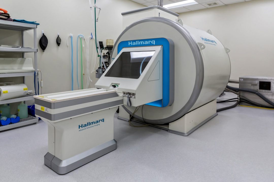
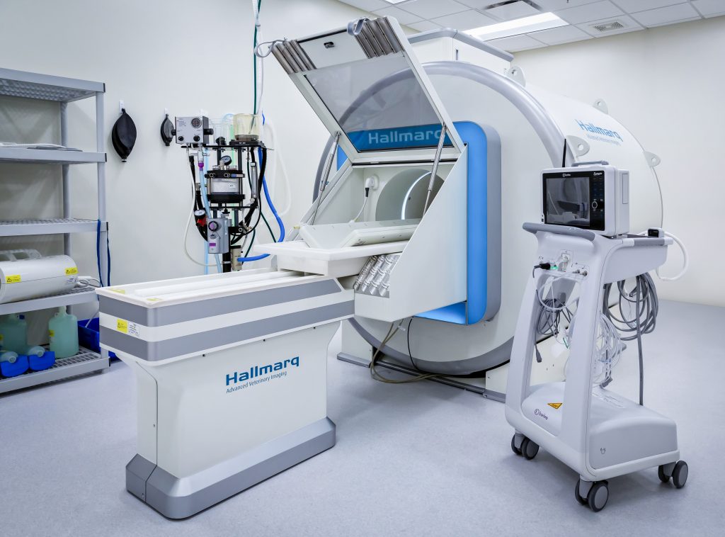
Let’s talk MIRA at Hallmarq
Hallmarq Veterinary Imaging has over two decades of experience in the design and manufacture of MRI machines. Their MIRA 1.5T small animal MRI considers the fact that animal anatomy is very different to that of humans! MIRA incorporates advanced features specifically designed to enhance imaging quality and diagnostic precision for veterinary applications.
Enhanced RF Coil Coverage
MIRA offers superior image quality thanks to its dual coil system, which significantly boosts signal intensity across larger body regions. This enhanced coverage allows for faster scans while maintaining high-resolution images, making it ideal for imaging a wide variety of pets, from the smallest of Chihuahuas to the largest of Great Danes.
Extended V-Shaped Phased Array Spine Coil:
The system includes a specially designed V-shaped phased array spine coil, which, when combined with an 8-channel flexible coil, increases scan coverage for larger animals. This feature is particularly beneficial for scanning difficult-to-image regions, such as the brachial plexus (or shoulder), providing greater clarity and detail.
Veterinary-Optimized MRI Software:
MIRA is equipped with cutting-edge veterinary-optimised software that delivers exceptional high-resolution detail. This software includes features such as pre-saturation and fat suppression for clearer images, blood flow compensation to reduce motion artifacts, single-shot fast spin echo for rapid imaging, and intensity correction. Additionally, it supports diffusion imaging with automatic ADC maps, which are crucial for diagnosing neurological conditions by detecting tissue changes with high precision.
Built-in Radiofrequency (RF) Shield:
MIRA has a built-in RF shield which makes it far more accessible for veterinary practices looking to offer small animal 1.5T at their hospital. There is no need for a purpose-built, shielded, room, keeping installation costs and complexities to a minimum. If more practices are able to offer MRI for your cat or dog, then more patients can reap the benefits of the gold standard in diagnostic imaging.
Hallmarq’s 1.5T high-field magnet with its own shielding is revolutionary. It provides us with a lot more flexibility to look at our patients in greater detail.
Dr. Christiane Massicotte, DVM, MS, PhD, DACVIM (Neurology), Veterinary Care & Speciality Group, Chattanooga, TN
Zero-helium MRI from MIRA
Hallmarq has designed MIRA to be 100% helium-free. A first in the veterinary industry, it means that your vet won’t have to spend valuable time and money on sourcing a finite resource. Instead, the machine remains available for uninterrupted work flow meaning that patients can be seen quickly and a diagnosis and care plan put into place. It also means we’re looking out for the environment too, so it’s a win-win for your pet and the planet!
These advanced features make MIRA MRI a powerful tool in providing accurate and efficient diagnostic imaging for veterinary patients.
Where can my pet access advanced imaging?
Hallmarq has partnered with customers since 2000 to support over half a million animal scans in 27 countries around the globe. With over 160 installations, explore the map to find your nearest Hallmarq system. If your veterinarian doesn’t yet offer MRI at their practice, then ask for a referral to your nearest Hallmarq small animal MRI site.
Hallmarq’s 1.5T MRI is designed specifically for small animal anatomy and delivers accurate and detailed images with great image resolution and contrast. The essential safety features and operator friendly software make it an asset to any veterinary neurologist.
Dr. Franziska Fitz, DVM, GPCertSAS, DACVIM (Neurology/Neurosurgery), Las Vegas Veterinary Speciality Center
At Hallmarq, our mission is to improve the lives of animals and those who care for them by increasing access to advanced imaging technology through awareness, education, and affordable and safe veterinary products. With over two decades of veterinary-specific experience in the design and manufacture of MRI and CT machines for dogs, cats and horses, we speak vet at hallmarq.net.
ARE YOU INTERESTED IN VISIONARY VETERINARY IMAGING?