

When your horse goes lame, you want answers, and fast. But diagnosing the cause of lameness isn’t always straightforward. The process often takes time and involves several steps before reaching a likely diagnosis and, ultimately, a treatment plan. Our step-by-step guide covers all you need to know about equine lameness diagnosis and what you can expect when looking to treat your lame horse.
You can also find out more on our Talk Lameness website. Specifically designed as a resource for horse owners, Talk Lameness walks you through the signs of horse lameness, what to look out for, how to record and note what you see, common symptoms and lot’s more. With a lameness quiz to test your knowledge and downloadable pdfs for you to print off and keep, Talk Lameness is a one-stop-shop for your lameness learning!
The lameness workup: what’s typically involved
A thorough clinical examination is usually the most important starting point. This is followed by nerve blocks or joint blocks to help localise the source of pain. Once the area(s) of concern have been identified, imaging is the next step. Radiographs (X-rays) and ultrasound are typically used first but, if the findings don’t fully explain the degree of lameness, more advanced imaging may be required. These techniques, such as Magnetic Resonance Imaging (MRI), Computed Tomography (CT) and Positron Emission Tomography (PET) provide detailed three-dimensional images that can reveal issues not visible with standard methods.
The lameness workup may sound like a long process, but it’s truly necessary. Taking each step carefully not only leads to more accurate answers, it can also help avoid unnecessary costs and reduce stress for both horse and owner.
Which imaging modality for your lame horse?
First-line imaging modalities, such as radiographs (X-rays) and ultrasound, are performed with the horse awake. In most cases, a small amount of sedation is used to make the procedure quicker and less stressful for everyone. When more advanced imaging is needed, however, there are several options. Both MRI and CT scans can be carried out with the horse either standing, or under general anaesthesia.
Different types of advanced imaging machines provide varying levels of detail and your vet will guide you towards the most suitable choice for your horse based on the findings so far. Factors such as temperament and age of the horse and available budget also come into play.
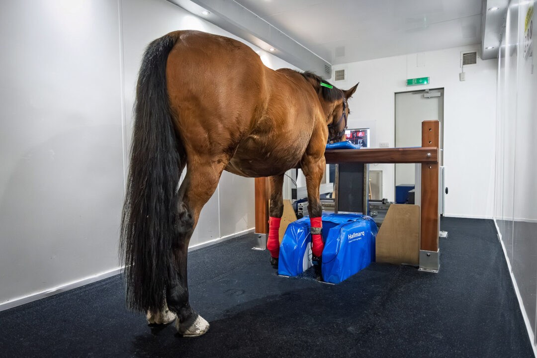
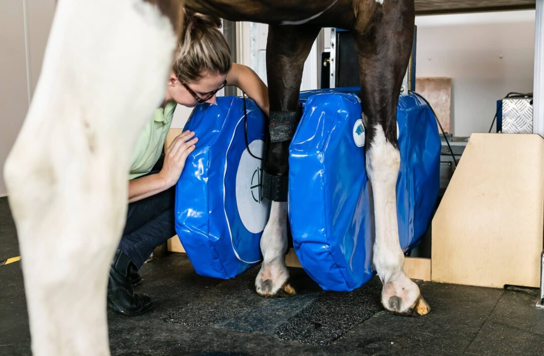
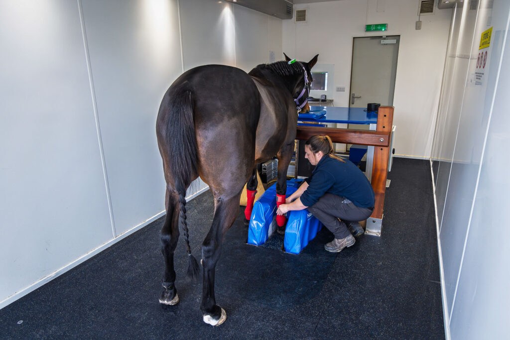
When it comes to standing advanced imaging, Hallmarq’s Standing Equine MRI has set the standard in image quality and safety. By allowing horses to be scanned while standing under mild sedation, and without the risks associated with general anaesthesia, Hallmarq technology provides veterinarians with the clear, detailed images needed to diagnose complex lameness cases and begin treatment sooner.
Let’s take a closer look at the key steps in this process – from block, to image, to treatment – and how each stage contributes to a faster, clearer diagnosis for your horse.
Step 1: clinical examination
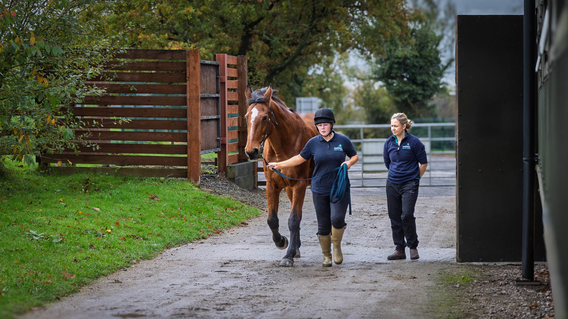
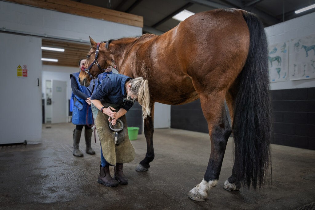
The journey to a sound diagnosis always begins with a thorough clinical examination to assess movement pattern and lameness scale. Before any nerve blocks or imaging are considered, your vet will carefully consider your horse both at rest and in motion, for example with flexion tests. This usually includes watching the horse walk and trot in a straight line, on the lunge and sometimes in canter and under saddle, depending on the case.
Your vet will palpate the limbs with precision, checking for areas of heat, swelling or sensitivity that may indicate inflammation or injury. Flexion tests may be performed to highlight pain in specific areas.
Observing the horse in motion provides valuable insight into stride length, rhythm and whether lameness is more noticeable on a straight line or a circle. This is routinely done by lunging your horse in a circle where the vet can note any subtle changes in gait, posture or symmetry. Assessing your horse on lunge offers the first clues as to where the problem may lie.
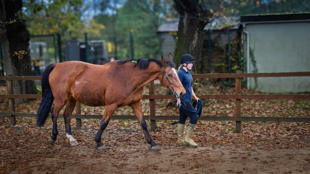
This comprehensive first step forms the foundation of every lameness assessment. It establishes a baseline understanding of how your horse is moving on the day of the exam and provides the context needed to guide the next stage of investigation. Without it, even the most advanced diagnostic tools risk being used without focus or efficiency. In other words, a careful physical exam is not just routine, it’s an essential starting point for accurate, timely diagnosis.
Step 2: Nerve blocks and intrasynovial anaesthesia
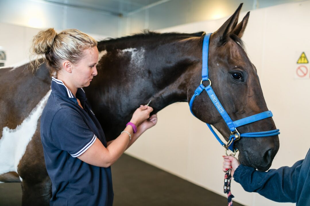
Once the lame limb (or limbs) has been identified the next logical step in the lameness work-up is the use of diagnostic nerve and joint blocks. These involve carefully injecting a small amount of local anaesthetic into targeted areas of the limb. The injection can be placed either within synovial structures (such as joints, bursae or tendon sheaths) or subcutaneously close to specific nerves. The effect is temporary but powerful: by numbing sensation in a specific region, your vet can observe whether the lameness changes; improving, worsening or even appearing to switch limbs.
The process is systematic and methodical. Unless the clinical exam has already highlighted an area, your vet will usually begin at the lowest point of the limb, such as the foot and then work gradually upwards. After each block, the horse’s movement is reassessed. If the lameness is abolished or improves significantly – or if it appears to switch limbs – it strongly suggests the pain originates in that region. If not, the vet continues higher until the affected area is identified.
For horse owners, it can be useful to think of nerve blocks as flipping off light switches in different rooms of a house. By turning them off one by one, you can quickly find out where the problem lies. This precise localisation is invaluable: it narrows down the possibilities, ensures imaging is focused on the right area, and helps avoid unnecessary or inconclusive diagnostics.
However, not all cases are straightforward. Horses may have multiple areas of pain in the same limb or in different limbs. In addition, nerve blocks are not perfectly specific; the local anaesthetic can spread between synovial structures or further up the limb, which can complicate interpretation. Certain pathologies are also trickier to block accurately. Like any procedure, there are potential risks and occasional adverse reactions, though these are relatively rare.
Overall, diagnostic blocks are safe, widely used and form an essential bridge between the clinical exam and advanced imaging techniques such as standing equine MRI. They save time, reduce costs and, most importantly, give your vet the confidence to investigate the right part of the limb in detail.
Step 3: First-line imaging modalities (radiographs and ultrasound)
Once the likely area of pain has been identified through the clinical exam and nerve blocks, the next stage in the lameness investigation is usually diagnostic imaging with tools that are widely available in equine practice. The two most common starting points are radiographs and ultrasound, each with its own advantages and limitations.
Radiography (X-ray) is often the primary method of assessing bone. It’s relatively inexpensive and can often be performed in the field without bringing the horse into the clinic. X-rays can reveal changes such as osteoarthritis, fractures, osseous cyst-like lesions and changes of bone density. However, changes in bone density generally need to reach 30–50% before they become visible, which means that early pathology can be missed. Radiographs also produce two-dimensional images, where superimposition can obscure detail and they cannot provide information about surrounding soft tissues.
Ultrasound is widely used to image soft tissues in both human and veterinary medicine. It is cost-effective, but it is highly operator-dependent. Because ultrasound waves cannot pass through bone, only the bone’s surface and the adjacent soft tissues can be assessed. Despite this, ultrasonography is particularly valuable for evaluating tendons, ligaments and joint capsules and is great for assessing progression of soft tissue injuries over time.
In many cases, these imaging methods are enough to reach a diagnosis and plan treatment. However, they can miss subtle or early changes and may not provide a complete picture of complex structures. The hoof, in particular, is a challenging area due to the dense hoof capsule. When results are inconclusive, or when a more detailed picture is needed, the logical next step is to proceed to advanced imaging.
Hallmarq’s unique Standing Equine MRI system provides unparalleled detail of both bone and soft tissue in the lower limb in a lameness examination. By allowing horses to be scanned safely while standing under mild sedation, without the risks of general anaesthesia, it delivers diagnostic answers in over 90% [1] of lameness cases.
Step 4: Advanced imaging – Standing Equine MRI
When other diagnostic tests don’t provide a clear answer, MRI often does. Standing Equine MRI is one of veterinary medicine’s most valuable non-invasive imaging technologies, proving the diagnosis of clinical signs in over 90% of lameness cases. MRI is considered the gold standard for assessing soft tissue injuries and it is also extremely useful for identify bone pathology. MRI provides detailed information about a lesion’s severity and activity, helping guide both treatment and prognosis.

Unlike radiographs, which require a 30–50% change in bone density before lesions appear, MRI can detect early stage changes such as bone oedema and sclerosis. For soft tissue, MRI captures subtle injuries to ligaments and tendons that may be missed on ultrasound and provides greater detail through imaging in multiple directions and sequences. Hallmarq’s Standing Equine MRI is specifically designed for horses, allowing them to be scanned while standing under light sedation. By avoiding general anaesthesia, the process is safer, less stressful and more practical for both horse and owner.
MRI is particularly useful for conditions such as:
- Foot lameness (navicular apparatus and other structures within the hoof) – Standing MRI has revolutionised the diagnosis of conditions within the hoof capsule, enabling the evaluation of structures previously unseen with other modalities. What was once referred to broadly as “navicular disease” can now be differentiated into specific pathologies involving the deep digital flexor tendon (DDFT), navicular bone, navicular bursa, collateral sesamoidean ligament and distal impar ligament. Deep digital flexor tendinitis is the most common cause of foot pain and MRI provides the most accurate diagnosis. MRI also allows complete evaluation of the collateral ligaments of the coffin joint, whereas ultrasound could only visualise the proximal aspect.
- Soft tissue injuries higher up the limb – Injuries to the DDFT, SDFT, proximal suspensory ligament and suspensory branches can be evaluated in much greater detail with MRI. Unlike ultrasound, MRI produces slices in multiple directions and uses different sequences, allowing vets to distinguish between acute and chronic lesions and to assess their true extent.
- Read more about injury of the proximal metacarpal region here and about ultrasound vs MRI for proximal suspensory ligament injuries here.
- Assessing progression and healing – MRI is invaluable for monitoring recovery and guiding rehabilitation plans. Repeat scans can show whether a lesion is improving, stable or worsening, even when these changes are not obvious clinically or on other imaging modalities. This makes MRI particularly powerful for long-term management of complex injuries.
- Discover more about the assessment of fractures here and the value of rescans here.
- Solar penetrations – While radiographs can identify tracts and osseous involvement, they often fail to show the full extent of penetration. with up to 66% [2] of navicular bursa puncture cases appearing normal on X-rays. Ultrasonography is also limited by poor penetration through the hoof capsule. MRI, however, can visualise the entire tract and the structures affected, providing a much clearer assessment.
- Learn about chronic solar penetration here and the role of MRI in equine solar penetration injuries here.
- Bone oedema-like lesions – MRI is considered the gold standard for detecting bone oedema in live horses – an early indicator of bone response to loading or injury. These changes are invisible on radiographs but correlate strongly with clinical signs of pain. Detecting them early allows vets to adjust management before more severe pathology develops.
- Fractures and fracture healing – MRI can identify associated bone oedema around fractures and monitor the resolution of these changes on repeat scans, providing valuable information for prognosis and return-to-work planning.
Because the horse remains standing, the scan is not only safer but also more efficient. It eliminates the risks of anaesthesia, reduces recovery time and often lowers costs compared with traditional “down” MRI systems. Most importantly, with MRI diagnostic in over 90% of lameness cases, it remains one of the most powerful tools available to give vets, and owners, the answers they need.
Step 5: from imaging to treatment
The true value of advanced imaging lies not just in finding the problem, but in shaping what happens next. A scan on its own is only part of the story, MRI findings are always interpreted in the context of the full clinical picture: the lameness evaluation, your horse’s history and other diagnostic tests. Looked at together, these pieces become a powerful decision-making tool. This holistic approach allows your vet to move from uncertainty to a clear, actionable plan.
With the precise information that Magnetic Resonance Imaging provides, your vet can:
- Design a tailored rehabilitation plan that adapts to your horse’s individual needs, its progress and the degree of healing shown on repeat MRI scans.
- Recommend medication or surgery where appropriate, based on accurate localisation and assessment of the lesion.
- Advise on correct shoeing. Knowing exactly which structures are affected, and to what degree, allows for the ideal shoeing strategy to improve your horse’s comfort and support healing.
- Give a clearer prognosis. Understanding the nature and severity of the injury helps you plan realistically for your horse’s future, whether that means a return to competition, a modified workload or long-term management.
This is where Hallmarq’s Standing Equine MRI proves its worth. The high-quality images produced by our system give vets the confidence to recommend the right treatment from the outset, avoiding unnecessary delays and reducing the risk of recurrence. In short, moving from image to treatment with clarity means a faster return to soundness and a better long-term outlook for your horse.
Final thoughts
Lameness can be one of the most frustrating challenges for both horse and owner. What matters most is not just finding an answer, but finding the right one quickly and with confidence. By following a structured pathway, from the lameness examination, diagnostic analgesia, diagnostic imaging, equine vets can build a complete picture of the problem and map out the most effective route to recovery.
You can also find out more on our Talk Lameness website. Specifically designed as a resource for horse owners, Talk Lameness walks you through the signs of horse lameness, what to look out for, how to record and note what you see, common symptoms and lot’s more. With a lameness quiz to test your knowledge and downloadable pdfs for you to print off and keep, Talk Lameness is a one-stop-shop for your lameness learning!
Standing equine MRI has transformed this process. By offering detailed, three-dimensional insight without the risks of general anaesthetic, it bridges the gap between uncertainty and clarity. For owners, that means fewer unanswered questions and more informed decisions; for horses, it means targeted treatment that supports comfort, performance and long-term soundness.
The next time lameness becomes a concern, ask your vet not only what they can see, but how they plan to get there. With Hallmarq’s standing equine MRI as part of the diagnostic toolkit, the chances of reaching the right answer, and the right outcome, have never been stronger.
Find out more about Hallmarq Standing Equine MRI.
References
[1] Morgan, Jessica M., Helen Aceto, Timothy Manzi, and Elizabeth J. Davidson. ‘Incidence and Risk Factors for Complications Associated with Equine General Anaesthesia for Elective MAGNETIC RESONANCE IMAGING’.Equine Veterinary Journal, 7 November 2023, evj.14026. https://doi.org/10.1111/evj.14026.
[2] Meehan et al, ‘Should I use magnetic resonance imaging to evaluate horses with foot penetrations.’ Equine Veterinary Education, 26 January 2017, eve. 12724 https://beva.onlinelibrary.wiley.com/doi/10.1111/eve.12724