




Why understanding the pathology matters in proximal suspensory disease
Proximal suspensory disease (PSD) remains one of the most common causes of poor performance and lameness in sport horses. Although it can affect both the forelimbs and hindlimbs, it encompasses a spectrum of pathological changes in the area of the ligament origin. Injury may involve the suspensory ligament itself, its attachment to the proximal metacarpal or metatarsal bones (enthesopathy), adjacent bone pathology, or a combination. As our understanding of the condition evolved, it has become increasingly more apparent that horses presenting with similar clinical signs may have very different underlying pathology.
For horses with chronic proximal suspensory pain, particularly in the hindlimb, plantar fasciotomy combined with deep branch lateral plantar neurectomy has become a well-established surgical treatment option with encouraging reported outcomes in appropriately selected cases (crucial for the success). When a horse blocks to the proximal suspensory region and surgery is already being considered, it is fair to ask: why not proceed straight to surgery?
For Anna Drahonovska, DVM, MRCVS, the answer lies in recognising that not every horse with proximal suspensory pain has the same underlying pathology, stating:
Identifying the structures involved and the extent of the injury is an important part of deciding whether surgery is the most appropriate treatment.Ultimately, the success of the procedure, the rehabilitation programme and the horse’s prognosis all depend on understanding exactly what pathology is present.”
Anna Drahonovska DVM MRCVS
Understanding Plantar Fasciotomy and Neurectomy
How does Plantar Fasciotomy and Neurectomy work?
Plantar fasciotomy and neurectomy (PFN) has become an established surgical treatment for selected horses with chronic hindlimb proximal suspensory desmitis that fail to respond to appropriate conservative management and have suitable limb conformation, without advanced secondary pathological changes (Dyson 2012).
The procedure combines decompression of the proximal suspensory region by releasing the plantar fascia with neurectomy of the deep branch of the lateral plantar nerve. Reported return-to-performance rates are generally between 70 and 85% in appropriately selected horses, although outcomes depend on accurate case selection, concurrent orthopaedic disease, limb conformation and chronicity of injury.
- Plantar fasciotomy: involves releasing the deep plantar fascia overlying the proximal suspensory ligament. Chronic injury may be associated with increased pressure within this relatively non-compliant fascial compartment, and releasing the fascia is thought to improve the local mechanical environment for healing.
- Neurectomy: involves removing a small section of the deep branch of the lateral plantar nerve, reducing pain originating from the proximal suspensory ligament while largely preserving distal limb motor function. Although pain relief is considered the principal mechanism, the exact biological effects remain incompletely understood, and both denervation and mechanical decompression are likely to contribute to the clinical outcome.
Which horses are suitable candidates?
Current indications include chronic hindlimb PSLD confirmed by clinical examination, diagnostic analgesia, and imaging, particularly in horses that have failed after prolonged rehabilitation, controlled exercise, extracorporeal shockwave therapy, regenerative therapies, or other conservative treatments.
PFN is most commonly performed in sport horses expected to return to athletic performance. Appropriate case selection is essential. Horses with advanced suspensory degeneration, extensive osseous pathology, significant concurrent orthopaedic disease or unsuitable limb conformation may have less favourable outcomes.
For example, a horse with fissure of the proximal third metatarsal bone identifies on MRI would not be an appropriate candidate for plantar fasciotomy and neurectomy.
What outcomes can be expected?
Published studies generally report good to excellent results following PFN in appropriately selected horses. Approximately 70–85% of horses return to their previous or intended level of work, although reported success rates vary according to case selection, discipline, duration of injury, and criteria used to define success.
Complications are uncommon but may include persistent lameness, incomplete return to performance, neuroma formation, or recurrence of clinical signs. Long-term follow-up suggests that many horses maintain improved athletic function for several years after surgery.
Proximal suspensory disease: an umbrella term
Although proximal suspensory disease (PSD) is often referred to as a single condition, it encompasses a wide spectrum of pathological changes. While horses may present with similar clinical signs or localise pain to the proximal suspensory region, the pathology responsible for those signs can vary considerably. Injury may be confined to the suspensory ligament itself, involve its attachment to the proximal metacarpal or metatarsal bone (enthesis), the adjacent bone, or a combination of these.
The structures involved in the pathology are clinically important because they may influence treatment selection, expected outcome and rehabilitation requirement. Some horses have pathology largely confined to the ligament, while others have significant osseous involvement, including sclerosis, bone oedema, fissures or small avulsion fragments. Changes at the suspensory ligament origin (enthesopathy), where the ligament attaches to the bone, as well as concurrent pathology affecting adjacent structures, may also be present.
The term enthesopathy is used to describe any disorder affecting an enthesis—the site where a tendon, ligament, joint capsule or fascia attaches to bone (Auer et al 2019). Although these horses may all localise pain to the proximal suspensory region, they may not require the same treatment, rehabilitation programme or have the same prognosis.
Advances in diagnostic imaging have transformed our understanding of proximal suspensory disease, demonstrating that bone and soft tissue pathology frequently coexist rather than occurring in isolation.
Chrysanthi Pitaouli DMV, MRCVS Cert AVP
As a result, clinicians are now able to better characterise the underlying pathology and appreciate how these differences may influence treatment decisions and outcome.
The challenge, therefore, is not simply confirming that pain originates from the proximal suspensory region but determining exactly which structures are involved and the extent of pathology present. This raises an important question: can conventional imaging always provide enough information?
Is conventional imaging enough before surgery?
The first step: localising the source of pain
A thorough lameness examination and diagnostic analgesia remain the foundation of investigating horses with suspected proximal suspensory disease. Once pain has been localised to the proximal suspensory region, the next step is to determine exactly what pathology is present.
However, several techniques have been described for blocking the proximal suspensory region, each with recognised advantages and limitations, none of them diminishes local anaesthetic diffusion to adjacent structures, that may complicate interpretation. For this reason, diagnostic analgesia should always be interpreted alongside imaging findings rather than in isolation.
The next step: conventional imaging
Radiography and ultrasonography are the first-line imaging modalities for investigating the proximal suspensory region and provide complementary information about the bone and soft tissues. In many horses, they provide sufficient information to guide treatment.
However, they cannot always fully characterise the osseous component of proximal suspensory disease, determine lesion activity, identify concurrent pathology or provide all the information required to select the most appropriate candidates for surgery. This is where MRI may provide valuable additional information.
Where can MRI add value before surgery?
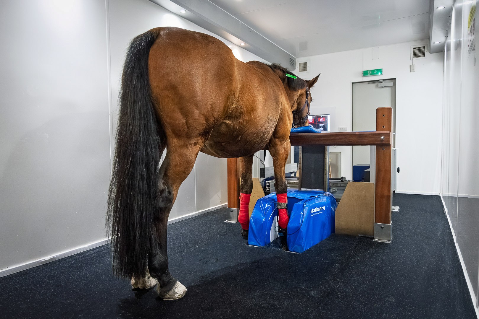
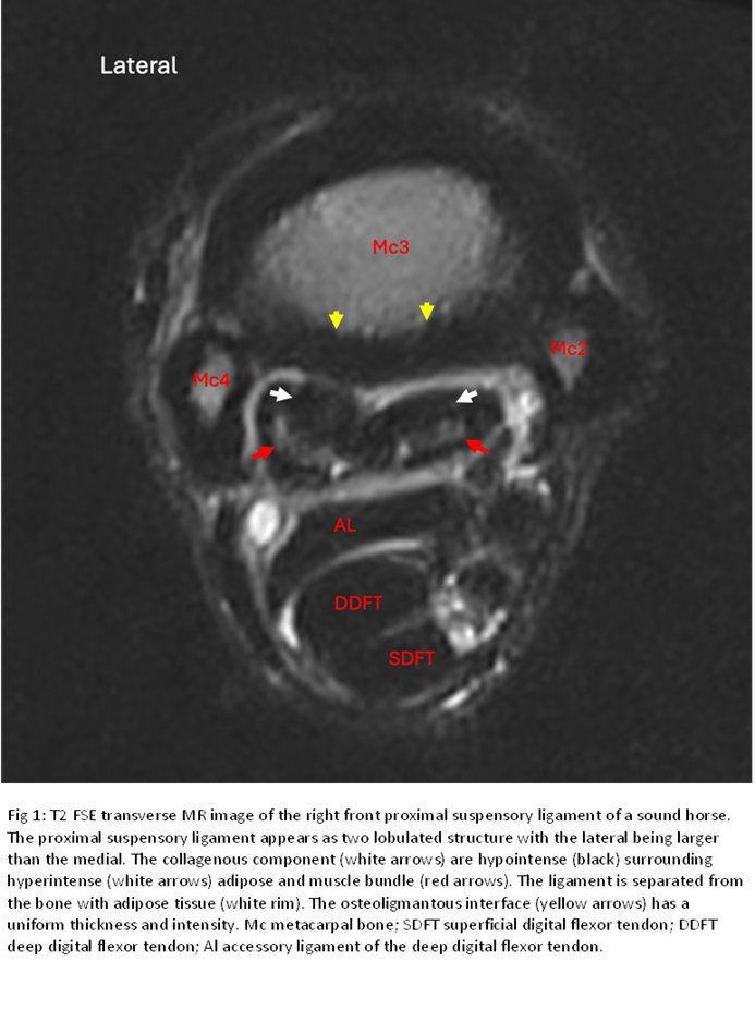
When surgery is already being considered, MRI provides information that goes beyond confirming the presence of proximal suspensory disease. By assessing both bone and soft tissue within a single examination, it allows clinicians to determine which structures are involved, the extent of pathology present and whether concurrent lesions may also be contributing to the horse’s clinical signs.
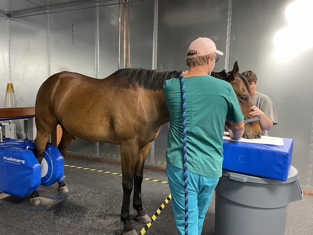
This additional information may influence whether surgery or which surgery is the most appropriate treatment, help refine the expected prognosis and guide postoperative rehabilitation. MRI may confirm that surgical intervention is appropriate, identify pathology that requires a modified treatment approach, or reveal concurrent lesions that could influence the expected outcome.
Rather than replacing clinical examination or conventional imaging, MRI complements the information already obtained, helping clinicians make more informed decisions before committing a horse to surgery. Ultimately, the aim is not to perform MRI before every horse undergoes surgery, but to ensure that horses are appropriately selected and follow up can be closely monitored. When performed, surgery should be the correct and most suitable intervention and any treatment decisions based on the best possible understanding of the underlying pathology.
Conclusion
The question is not whether every horse with proximal suspensory pain should undergo MRI before surgery. Rather, it is whether enough information has been gathered to make the best possible treatment decision for that individual patient/pathology.
Plantar fasciotomy and neurectomy remain well-established treatment options in appropriately selected cases and may subside to neurectomy only etc. However, because proximal suspensory disease encompasses a wide spectrum of pathological changes, understanding the structures involved and the extent of pathology is fundamental to selecting suitable surgical candidates, providing a realistic prognosis and developing an appropriate rehabilitation programme.
When uncertainty remains, MRI can provide valuable additional information to complement conventional investigation and help ensure that the most appropriate treatment is selected for the individual horse and that surgical intervention is undertaken only when the underlying pathology is likely to benefit from it.
References
- Hewet-Deadman C. Proximal suspensory desmopathy: current concepts in diagnosis and management. In Practice. (Review article/CPD publication.)
- Dyson SJ, Murray RC. 2012. Management of hindlimb proximal suspensory desmopathy by neurectomy of the deep branch of the lateral plantar nerve and plantar fasciotomy: 155 horses (2003–2008). Equine Veterinary Journal44:361–367.
- Tatarniuk DM, et al. 2021. Outcome following neurectomy of the deep branch lateral plantar nerve and plantar fasciotomy for hindlimb proximal suspensory desmopathy in western performance horses. Veterinary Surgery50:273–282.
- Scharf A, et al. 2022. Suspensory ligament size does not change after plantar fasciotomy and neurectomy of the deep branch of the lateral plantar nerve by ultrasonographic assessment. Veterinary Surgery 51:259–269.
- Auer JA, Stick JA, Kümmerle JM, Prange T (eds.). Equine Surgery. 5th ed. St. Louis, MO: Elsevier; 2019.