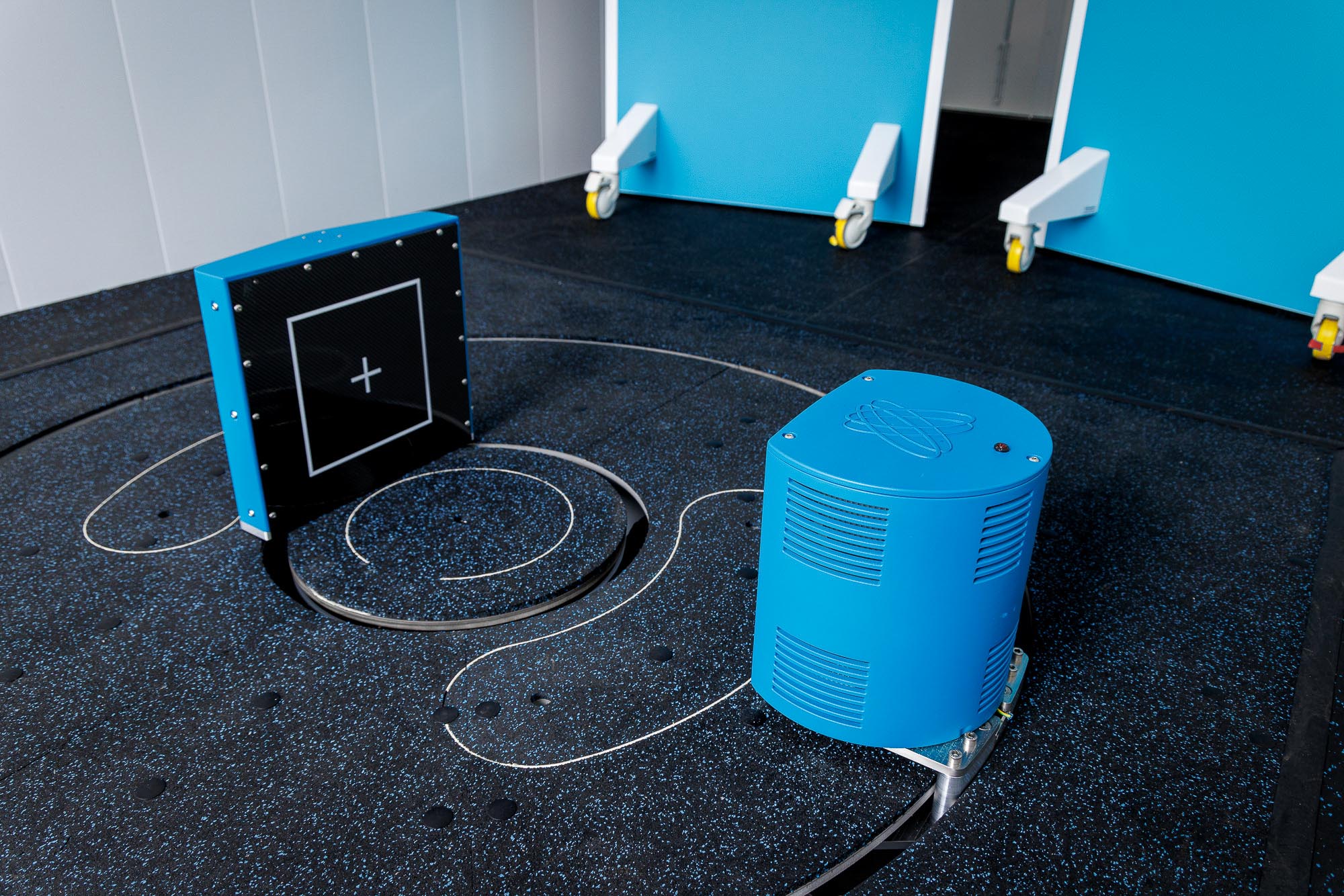


Did you know that Hallmarq’s Vision CT is being used by our customer sites globally, to help assess equine lower limb lameness? As a step up from DR – and complementary to Standing Equine MRI – this safe, effective, and affordable modality brings advanced 3D imaging to every equine practice.
But in which instances would you choose to use Vision CT? We asked some of our customers to share their clinical case studies to illustrate the value of Vision CT in aiding diagnosis. They make for fascinating reading and prove the versatility of the modality in everyday practice.
Did you know that Vision CT can…
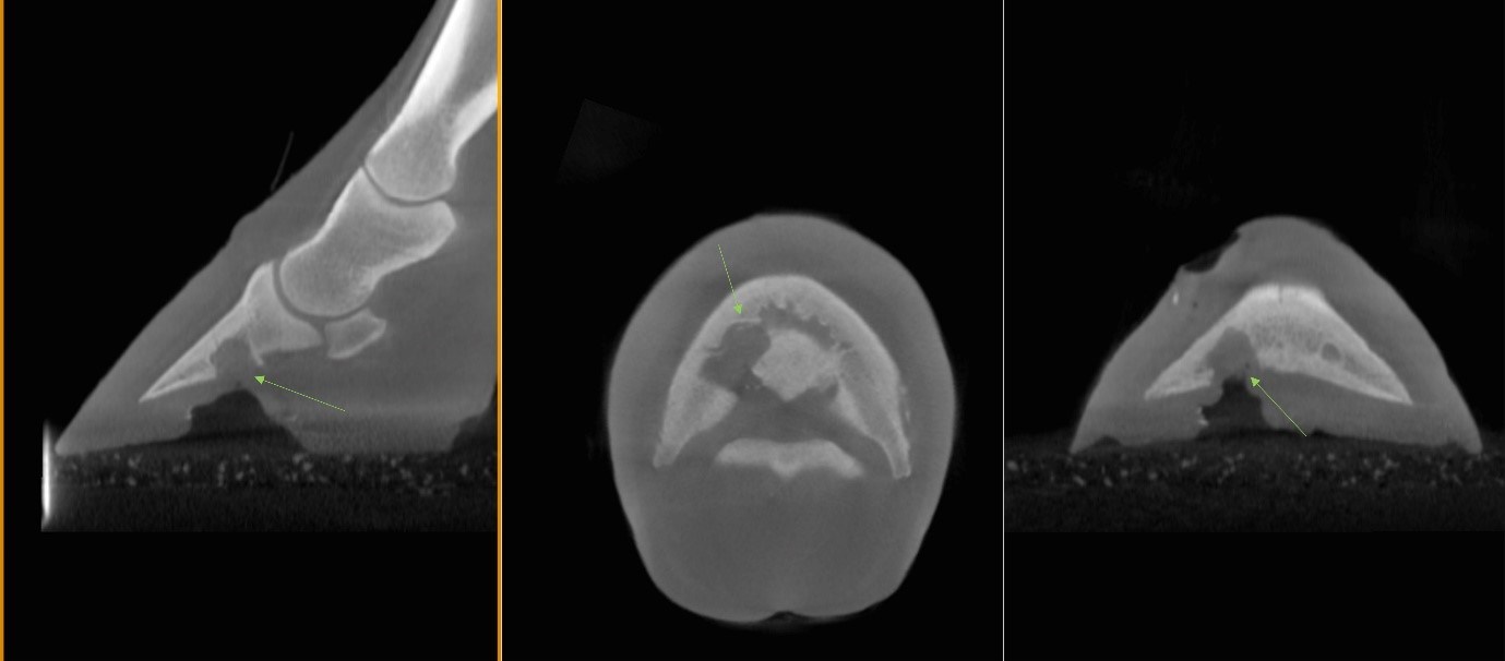
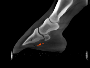
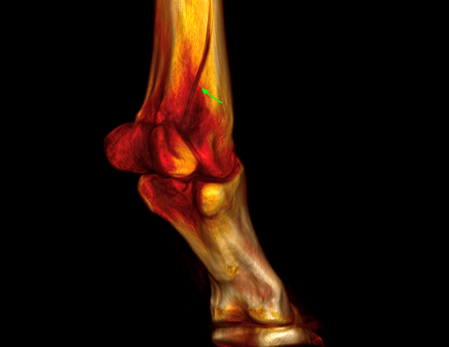
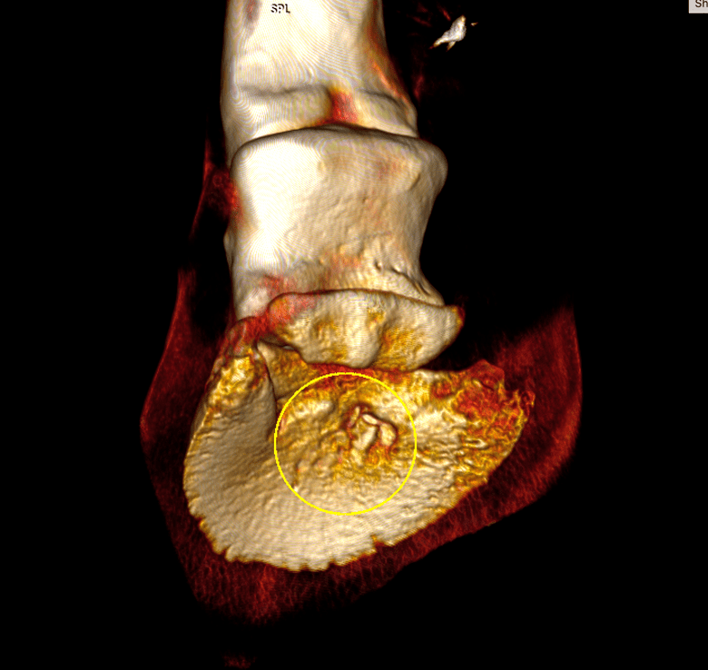
Aid in characterizing foreign bodies in the hoof and assist with surgical planning
Radiographs – performed on two occasions – suggested either a foreign body or a small bony fragment adjacent to the distal phalanx. Imaging with Vision CT however, revealed that what appeared to be a small bony fragment on radiographs was more consistent with metallic artefact.
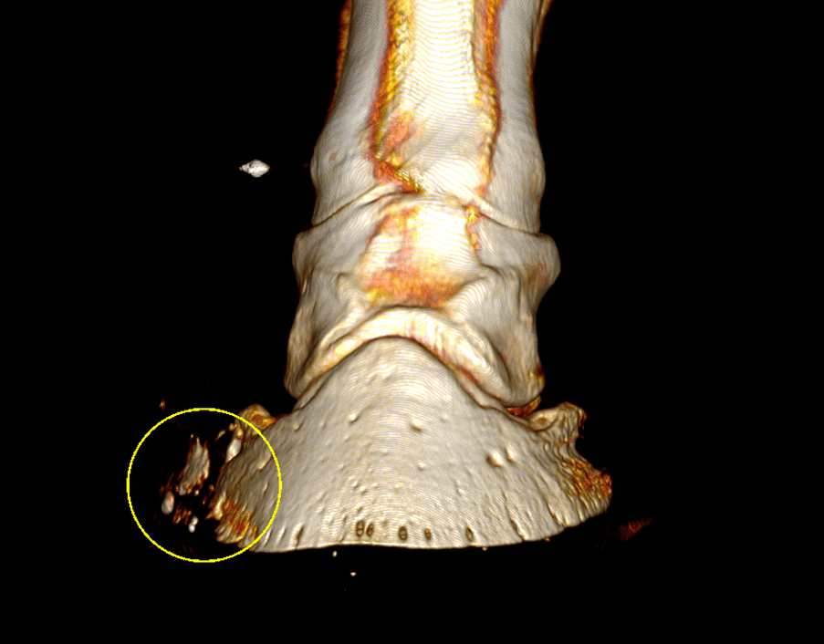
Provide the answers in cases involving metallic objects
Where metallic objects such as fragments from a rusty nail or surgical implants like screws are present, the diagnostic efficacy of MRI can be compromised. In this case, CT imaging provided additional structural data, proving instrumental in visualizing the tract and assessing the integrity of crucial internal foot structures.
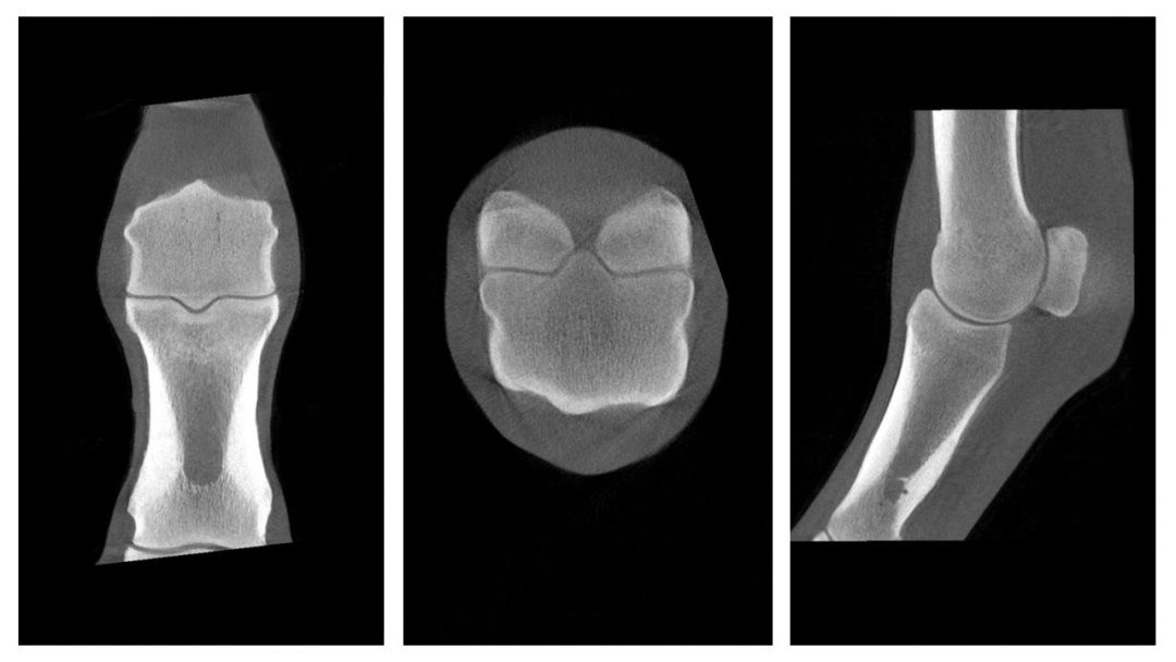
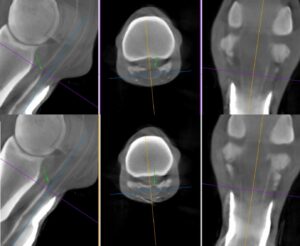
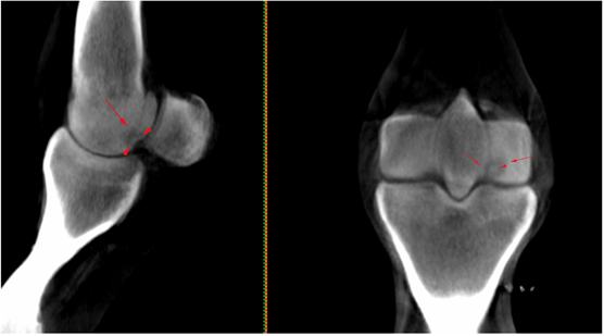
Deliver a more quantitative assessment of fracture lines
Imaging with Vision CT enabled a more quantitative and precise evaluation than the information provided by standard radiographs. CT conclusively confirmed the existence of a second non-displaced fracture line extending proximodistally through the axial aspect of the lateral proximal sesamoid bone and thus helped with surgical planning.
Complement Standing Equine MRI to help with pre-operative diagnosis
“Combining these advanced techniques has the potential to improve diagnostic sensitivity and specificity for these difficult injuries within the equine digital flexor tendon sheath.”
Dr Sarah Taylor (BVM&S MSc PhD Cert ES(Orth) DipECVS DipECVSMR MRCVS)
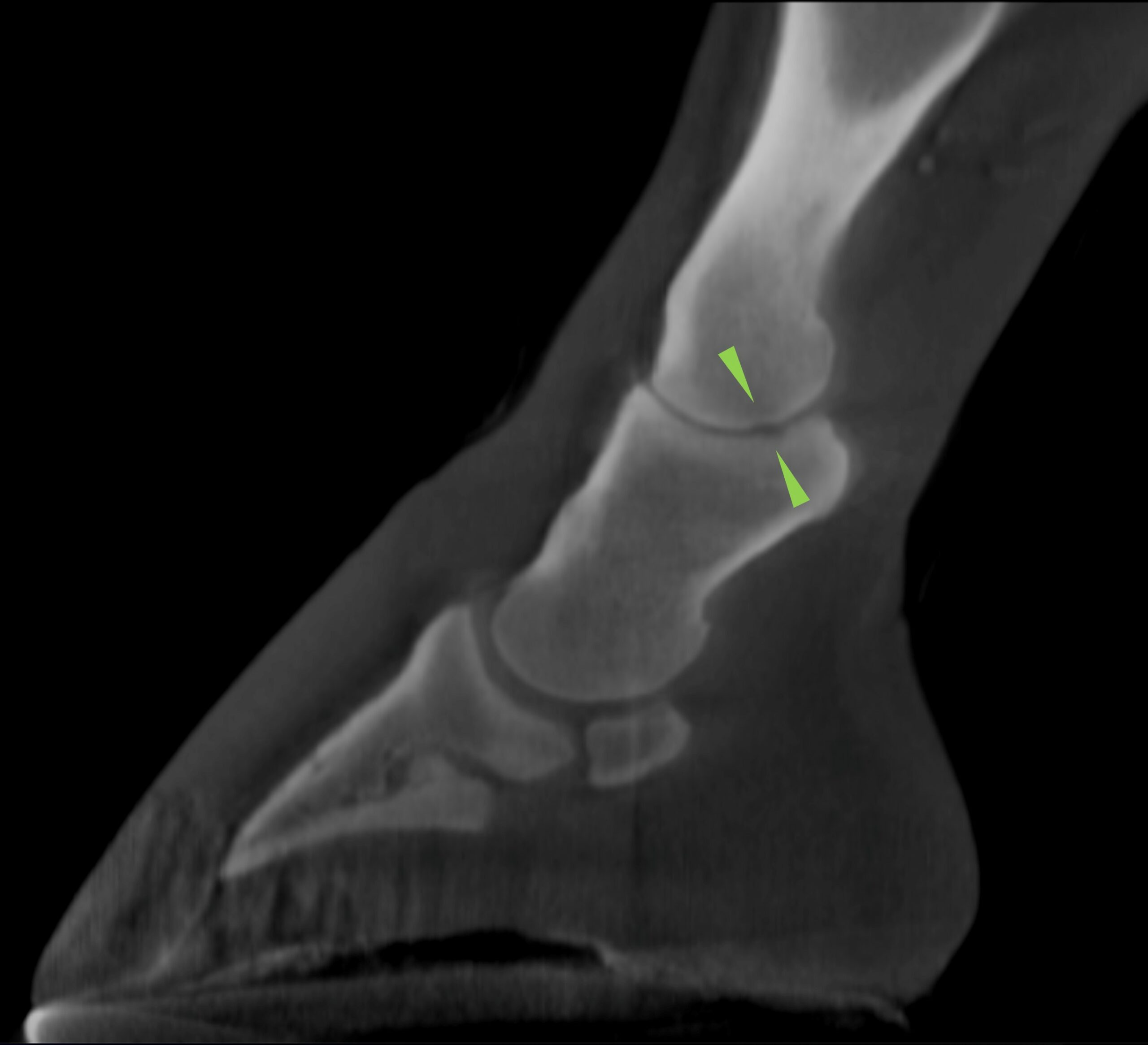
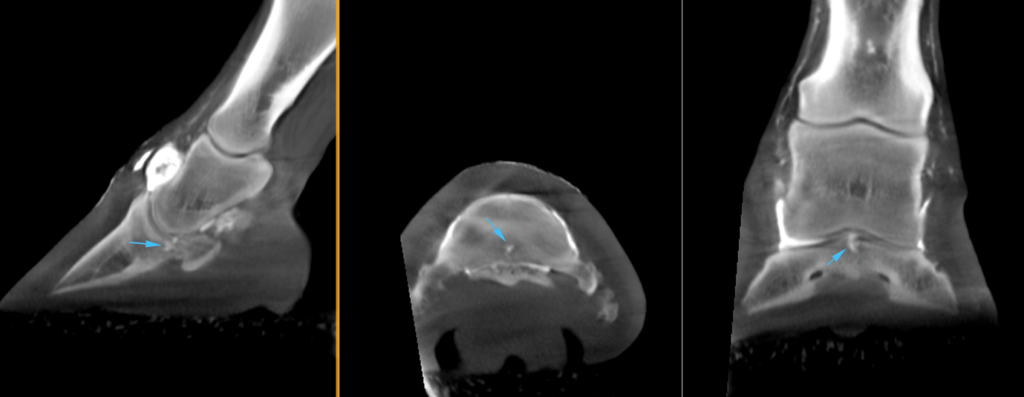
Provide a more quantitative assessment of lesions observed on radiographs
While radiographs showed lucency in the lateral condyle of the proximal phalanx, imaging with Vision CT enabled a more quantitative assessment, and revealed an elongated defect of the subchondral bone plate, at the lateral aspect of the pastern joint, involving the proximal and middle phalanges.
Aid in surgical planning for hoof debridement procedures
CT scans can be easily performed during and after surgery to assess the extent of debridement needed. In this particular case, repeat CT was performed 10 days after surgery to re-assess the pedal bone and ensure no further debridement was indicated.
Complement scintigraphic findings in diagnosing POD
In this case, the colt underwent scintigraphic imaging of the lumbar spine, pelvis, and hindlimbs to help identify the pathology present. The horse then underwent standing cone beam CT of the left hind fetlock joint and the conclusion made that the horse showed severe plantar osteochondral disease (POD).
Help better evaluate and characterize irregularities on the articular surface
Given the findings and suspicion of a region of focal disruption to the articular cartilage, a contrast-enhanced study was performed on the right front foot.
INTERESTED IN VISIONARY VETERINARY IMAGING?